General Information about Ashwagandha
Some preliminary studies have discovered that ashwagandha could have anti-cancer properties. It has been instructed that the herb has the potential to induce apoptosis (cell death) in most cancers cells and inhibit their progress. More analysis is required on this area, however the initial findings are promising.
However, whereas there have been numerous in vitro and animal studies supporting the consequences of ashwagandha, medical trials supporting its use are still restricted. In basic, analysis on herbal medication is limited, and the lack of regulations in the complement trade makes it challenging to determine the efficacy and security of merchandise.
One of the most significant benefits of ashwagandha is its potential to boost the immune system. Studies have proven that the herb can stimulate the manufacturing of white blood cells, which play an important function in preventing off infections and diseases. It additionally has anti-inflammatory properties that may help reduce irritation in the physique and help immune operate.
Conclusion
Ashwagandha is a powerful herb with potential therapeutic benefits for varied health points. While extra research is needed to determine its efficacy and safety absolutely, preliminary studies have proven promising outcomes. However, it is essential to consult with a healthcare skilled before incorporating any complement into one's routine, as it might interact with sure medicines and have adverse effects on some people. Additionally, it's crucial to purchase natural dietary supplements from respected sources to ensure their high quality and security. With correct research and caution, ashwagandha can be a wonderful addition to your well being and wellness regimen.
CNS (Central Nervous System) assist
Inflammation is a natural response of the body to struggle off infections and heal accidents. However, persistent inflammation can result in various illnesses and conditions like heart disease, diabetes, and arthritis. Ashwagandha has been found to have anti-inflammatory effects, which can assist in managing these situations. It has been proven to inhibit the manufacturing of pro-inflammatory cytokines and reduce markers of inflammation in the physique.
Immune System Support
Inflammatory Conditions
That being mentioned, let's take a look at some of the potential health advantages of ashwagandha which have been suggested by research so far.
In latest years, ashwagandha has gained reputation in the western world, and is now broadly obtainable in the form of dietary supplements. It is taken into account a natural different to medications for varied well being points, due to its potential therapeutic advantages.
Ashwagandha has been historically used as a nerve tonic to improve mind perform and cognitive abilities. Several research have shown that it might assist cut back anxiousness and stress, improve reminiscence and focus, and even promote higher sleep. It is believed to control stress hormones like cortisol within the physique, which may help handle stress and anxiety-related conditions.
Ashwagandha, also called Withania somnifera or Indian ginseng, is a powerful herb that has been utilized in Ayurvedic medicine for hundreds of years. It is a small shrub with yellow flowers and grows in dry areas of India, the Middle East, and components of Africa. Ashwagandha interprets to 'smell of horse' in Sanskrit, as a outcome of its strong smell and the idea that consuming it's going to give the power and stamina of a horse.
Anti-cancer Properties
Traditionally, ashwagandha has been used as an adaptogen, which is a substance that helps the body address stress and regulates the physiological processes to maintain homeostasis. It can additionally be known for its diuretic and sedative properties. In Ayurveda, ashwagandha is believed to balance the body's doshas (vata, pitta, and kapha) and promote general well being and longevity.
Resurfacing a distant flap from the trunk (abdominal skin flap according to Zoltan) is another option anxiety 37 weeks purchase ashwagandha 60 caps. To avoid these inconveniencies of a pedicled distant flap transfer, a free flap transfer can be performed. The choice between a pedicled and a free, microsurgically transplanted flap graft is determined by the localisation of the distal end point of the defect. The venous anastomosis is performed using the end-to-end technique with one of the two brachial veins. Based on the aesthetically reduced donor-site defects, the distally based medial upper arm flap (ulnar recurrent artery fasciocutaneous flap according to Maruyama) should be preferred over the distally based upper lateral arm flap according to Culbertson or the radial recurrent fasciocutaneous flap according to Maruyama. If no flap from the upper arm is possible, the pedicled radial artery flap according to Yang or pedicled ulnar artery flap according to Lovie are the next choice. In order to spare the radial artery, one should always check for the possibility of raising a proximal perforator flap. Through the abundance of musculature, this location is frequently used as a good recipient bed for replacement, and simple skin transplantation can frequently be performed here. Especially in the region of the middle third of the forearm and in the event of elective procedures carried out on suitable patients, the possibility of tissue expansion should be borne in mind. Larger defects can only be covered using the pedicled radial artery flap according to Yang or the pedicled ulnar artery flap according to Lovie. The same applies to the ulnar artery where it is even more important to avoid postoperative sensibility impairment of the ulnar nerve. On the ulnar side, there is one proximal option, namely the proximal, ulnar artery perforator fascia flap according to El-Khatib. Because of the multiple pedicled flap options for the treatment of, for example, devastating polyregional injuries, a free flap transfer is rarely indicated. If such a free flap is not an option, a distant flap from the trunk (abdominal skin flap according to Zoltan) is the last resort before considering amputation. A further important indication is to improve the positioning of constructions involving the extensor tendons. Larger defects are preferably covered with the posterior interosseous artery flap according to Penteado or Zancolli. For defects in the region of the dorsal wrist, however, care must be taken to ensure that the communicating branch of the posterior interosseous artery along with the anterior interosseous artery is preserved. If the posterior interosseous flap is not possible, further options include the distally based lateral antebrachial neurocutaneous flap according to Bertelli and the distally based medial antebrachial neurocutaneous flap according to Bertelli. In the presence of even larger defects, the further selection of flaps is made as discussed above. Since the dorsal side of the hand and the wrist are especially important for aesthetic reasons, one should take care not to use flap grafts that are too thick. If this proves to be unavoidable in the first instance, the first flap correction. Alternatively, thin pedicled fascia flap grafts can be used in combination with a moderately thick split-thickness skin transplant. With this technique, exceptional aesthetic results can be achieved, and only on rare occasions will postoperative corrections be necessary. According to Masquelet, four different regions can be distinguished: the dorsal, palmar, lateral and medial regions of the wrist. Since the distal region of the forearm is one of the low resistance zones, simple skin transplantation is generally only employed as a temporary coverage or as a last resort. For the coverage of smaller to moderately large defects, rotation flaps from the region of the lower arm can be used. Aside from the limitations imposed by the dimension of the flap, however, one must also consider the poor aesthetic results which, in particular, can be observed in the dorsal region of the lower arm. If the posterior interosseous flap is not available, further options include the distally based lateral antebrachial neurocutaneous flap according to Bertelli and the distally based medial antebrachial neurocutaneous flap according to Bertelli. However, one must keep in mind that raising these flaps will result in sensible impairment of the distal forearm. Larger defects can only be covered with the pedicled radial artery flap according to Yang or the pedicled ulnar artery flap according to Lovie. On the ulnar side there is one proximal option, namely the distal, ulnar artery perforator flap according to Becker. Because of multiple pedicled flap options, free flap transfer is rarely indicated, as. If a free flap is not available for such defects, a distant flap from the groin (groin flap according to McGregor) is the final option before considering amputation. A further and more frequent indication is observed in recurrent carpal tunnel syndromes, where simultaneous coverage and cushioning of the blood supply is necessary to protect the median nerve. Suitable options for cushioning and revascularisation of the median nerve primarily include the distal ulnar artery perforator flap according to Becker and the pronator quadratus muscle flap according to Dellon. For small to moderately large defects, the posterior interosseous artery flap is the first-choice therapy since no primary vessels need to be sacrificed. Due to the large donor-site defects, the distally based lateral antebrachial neurocutaneous flap according to Bertelli and the distally based medial antebrachial neurocutaneous flap according to Bertelli should be employed as a second choice. Defects in the region of the wrist are preferably covered using a posterior interosseous artery flap. Complex injuries with open fractures in the region of the wrist represent a contraindication for these flaps. Because of the large donor-site defects, the distally based lateral antebrachial neurocutaneous flap according to Bertelli and the distally based medial antebrachial neurocutaneous flap according to Bertelli should only be applied as a second choice.
Nonfoodborne Vibrio infections: an important cause of morbidity and mortality in the United States anxiety 5 see 4 feel generic ashwagandha 60 caps on line, 19972006. Vibrio fluvialis: an unusual enteric pathogen of increasing public health concern. Presented to: Monitoring and Evaluation Team, Child and Adolescent Health and Development. Complete genome sequence and comparative genome analysis of enteropathogenic Escherichia coli O127:H6 strain E2348/69. Clinical and immunologic characteristics of Vibrio cholerae O139 Bengal infection in North American volunteers. Vibrio cholerae O139 in Calcutta, 19921998: incidence, antibiograms, and genotypes. Cholera toxin structure, gene regulation and pathophysiological and immunological aspects. Vibrio cholerae produces a second enterotoxin which affects intestinal tight junctions. Toxin, toxin-coregulated pili and the toxR regulon are essential for Vibrio cholerae pathogenesis in humans. Antagonism toward the intestinal microbiota and its effect on Vibrio cholerae virulence. Zinc supplementation in children with cholera in Bangladesh: Randomised controlled trial. Severe diarrhea caused by cholera toxin-producing Vibrio cholerae serogroup O75 infections acquired in the southeastern United States. The role of Gulf Coast oysters harvested in warmer months in Vibrio vulnificus infections in the United States, 1988 1996. Diarrheagenic Escherichia coli in sub-Saharan Africa: status, uncertainties and necessities. Pathogens associated with persistent diarrhoea in children in low and middle income countries: systematic review. Atypical enteropathogenic Escherichia coli infection and prolonged diarrhea in children. Immunological cross-reactivity between a heat-labile enterotoxin(s) of Escherichia coli and subunits of Vibrio cholerae enterotoxin. Review on pathogenicity mechanism of enterotoxigenic Escherichia coli and vaccines against it. Weapons of mass destruction: virulence factors of the global killer enterotoxigenic Escherichia coli. Relative efficacy of blood, urine, rectal swab, bone-marrow, and rose-spot cultures for recovery of Salmonella typhi in typhoid fever. Outbreaks of enterotoxigenic Escherichia coli infection in American adults: a clinical and epidemiologic profile. Endemically acquired foodborne outbreak of enterotoxin-producing Escherichia coli serotype O169:H41. Outbreak of enterotoxigenic Escherichia coli infection with an unusually long duration of illness. Humoral immune response to the heat-labile enterotoxin of Escherichia coli in naturally acquired diarrhea and antitoxin determination by passive immune hemolysis. Prevalence of enteric pathogens among international travelers with diarrhea acquired in Kenya (Mombasa), India (Goa), or Jamaica (Montego Bay). Preliminary incidence and trends of infections with pathogens transmitted commonly through food -foodborne diseases active surveillance network, 10 U. Preventing household transmission of Shiga toxin-producing Escherichia coli O157 infection: promptly separating siblings might be the key. A systematic review of vaccinations to reduce the shedding of Escherichia coli O157 in the faeces of domestic ruminants. Characterization of Saa, a novel autoagglutinating adhesin produced by locus of enterocyte effacement-negative Shiga-toxigenic Escherichia coli strains that are virulent for humans. Infections with verotoxin-producing Escherichia coli O157:H7 and other serotypes, including the outbreak strain O104:H4. The epidemiology and clinical aspects of the hemolytic-uremic syndrome in Minnesota. Infections in pediatric postdiarrheal hemolytic uremic syndrome: factors associated with identifying shiga toxin-producing Escherichia coli. Epidemic profile of Shigatoxin-producing Escherichia coli O104:H4 outbreak in Germany. Escherichia coli O157:H7 and the hemolytic-uremic syndrome: importance of early cultures in establishing the etiology. Causative species and serotypes of shigellosis in mainland China: systematic review and meta-analysis. Presumptive shigellosis: clinical and laboratory characteristics of Bangladeshi patients. Epidemiologic and clinical features of patients infected with Shigella who attended a diarrheal disease hospital in Bangladesh. Gastrointestinal and extra-intestinal manifestations of childhood shigellosis in a region where all four species of Shigella are endemic. Central nervous system manifestations of childhood shigellosis: prevalence, risk factors, and outcome. Low risk of hemolytic uremic syndrome after early effective antimicrobial therapy for Shigella dysenteriae type 1 infection in Bangladesh. Zinc supplementation in the management of shigellosis in malnourished children in Bangladesh. Green banana reduces clinical severity of childhood shigellosis: a double-blind, randomized, controlled clinical trial.
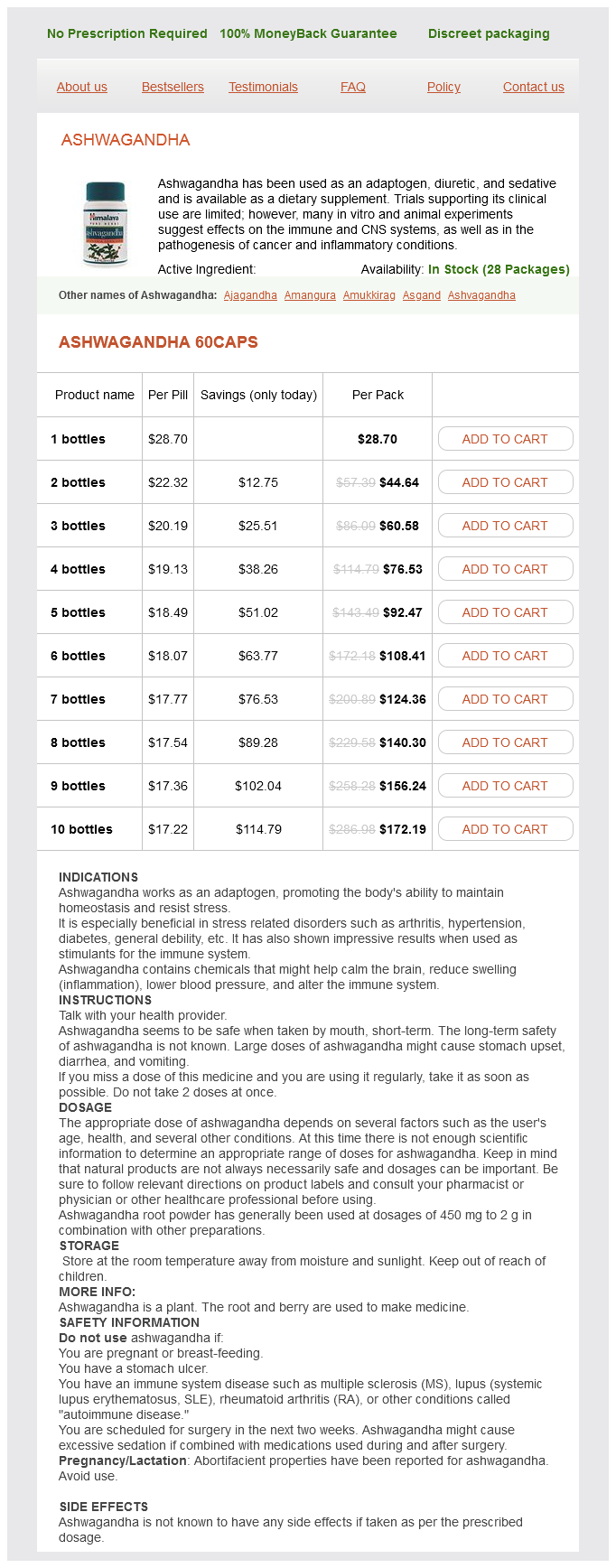
Ashwagandha Dosage and Price
Ashwagandha 60caps
- 1 bottles - $28.70
- 2 bottles - $44.64
- 3 bottles - $60.58
- 4 bottles - $76.53
- 5 bottles - $92.47
- 6 bottles - $108.41
- 7 bottles - $124.36
- 8 bottles - $140.30
- 9 bottles - $156.24
- 10 bottles - $172.19
This layer of connective tissue anxiety symptoms 10 year old order discount ashwagandha on line, whose firmness increases from proximal to distal, separates the radial compartment from the flexor lodge. It is attached proximally to the periosteum and distally to the attachment of the pronator quadratus muscle. The flap is centred on the course of the radial artery on the palmar side of the lower arm according to the requirements of the defect which has to 5. Subsequently, intensive physical therapy of the entire extremity as well as careful skin care in the transplant area are advised. In addition to the requirement on the thickness of the flap and the length of the vessel pedicle, functional and aesthetic aspects must be considered. In order to avoid postoperative contractures the flap may not exceed the joint folds proximally and distally. To improve the aesthetic outcome of the donor site defect, the skin of the radial forearm side must remain untouched. In addition, hair growth in this area might disturb the aesthetic result in the recipient site. In the area of the distal forearm the skin-fascia portion is thin with only little subcutaneous fat. A sufficient length of the vessel pedicle with distal pediculation can be obtained by raising the flap in the middle third of the forearm. After the planning of the flap is complete, the radial artery is identified on the proximal edge of the flap and in the wrist, encircled and retracted. Subcutaneous skin veins are ligatured, and one or two larger veins are exposed and prepared for a possible venous augmentations anastomosis. The medial and lateral cutaneous nerves of the forearm entering on the proximal flap edge will also be exposed and spared. As soon as the intermuscular septum between the flexor carpi radialis muscle on the ulnar side and the brachioradialis muscle on the radial side is presented, the tourniquet is released. With satisfactory perfusion, the radial artery and veins are ligatured proximally, and the muscular septum together with the flap is then sharply divided from the radius from proximal to distal, using magnifying loupes. Attention must be paid to obtain a complete haemostasis of the vessels supplying the brachioradialis, flexor carpi radialis, flexor pollicis longus and flexor digitorum superficialis muscles as well as the periosteal branches. Alternatively, a vascularised fasciocutaneous strip can be retained in the area of the vascular pedicle and sutured in place after a skin incision has been made to avoid compression of the pedicle. To improve venous drainage, a venous anastomosis between a superficial flap vein and a dorsal skin vein in end-to-end or end-to-side technique is recommended. The reconstruction of the radial artery by an inverted vein graft from the lower arm, usually the cephalic vein, is no longer recommended. To improve the transplant position the muscle bellies of the brachioradialis and flexor carpi radialis muscles should be approximated and the distal parts of the muscle belly of the flexor carpi ulnaris muscle should be transposed over the flexor tendons. Postoperative immobilisation of the forearm for 7 to 10 days is neces- Variants Radial artery fascia flap Over a curved skin incision, and using the same operation technique, the lower arm fascia alone is raised from the radial artery and veins. Care must be taken to leave the superficial side of the fascia covered with a thin layer of adipose tissue in order to prevent injury to the local capillary network. The vascularised fascia-fat flap is covered with a medium splitthickness skin transplant on the back of the hand, and with a full-thickness skin transplant in the palm. An improved healing rate of the skin transplant can be achieved if the covering takes place after 3 to 5 days (multiple-stage skin grafting procedure). Fascio-myocutaneous composite radial artery flap Because of the constant supply to the brachioradialis and radial flexor carpi muscles, both can be raised partially or completely with a fasciocutaneous flap at the radial vessels. On account of its close contact with the lower arm fascia the same applies to the tendon of the palmaris longus muscle. Osteo-fasciocutaneous composite radial artery flap As in the operation technique described above, the lateral intermuscular septum, with its attachment to the radius, is identified. Along its connection between the insertion of the pronator muscle (proximally) and the insertion of the brachioradialis muscle (distally) a corticocancellous vascularised radius bone transplant can be raised. To reduce fracture risk, attention must be paid that no more than half of the circumference of the radius is taken. For the same reason, a drill-hole should be made at the proximal and distal ends of the strip. The bone transplant is harvested using an oscillating saw, sparing the supplying vessels. Because of the unsatisfactory sensitivity, an additional operation is necessary to restore sensitivity in the neo-fingertip area. The fasciocutaneous flap with vascularised tendon components is indicated for the reconstruction of combined skin-tendon defects, especially on the side of the dorsum of the hand. The osteo-fasciocutaneous variant is suitable for the coverage of a combined skin-bone defect in the area of the middle hand and basic phalanx (P1). Contraindications to the radial artery flap are circulation disorders in the hand area and current injuries in the area of the anatomical snuff box. Although the application ofdistally pedicled radial artery flaps has been reported in cases where both palmar arches were missing, the radial artery flap should not be applied defects in patients with a negative Allen test, loss of flow reversal in Doppler sonography or absence of the palmar arch in the angiogram of the radial artery flap since the risk of donor site defects is increased. Especially in adipose patients with a thick subcutaneous fat layer in the lower arm area, the radial artery flap should be considered as a second choice therapy because of the large aesthetic donor site defect. With menopausal women the harvesting of a vascularised radius strip is also contraindicated on account of to the increased fracture risk. As most of the proximal perforators emerge 5 cm distally to the origin of the radial artery, the pivot point is located 5 cm below the intercondylar line.