General Information about Ceftin
Ceftin is used for treating bacterial infections, particularly these of the sinus, skin, lung, urinary tract, ear, and throat. These kinds of infections can be attributable to a variety of micro organism, similar to streptococcus, staphylococcus, and Hemophilus influenzae. Ceftin works by stopping the expansion and multiplication of these bacteria, thus serving to the physique's natural defense mechanisms to fight off the an infection.
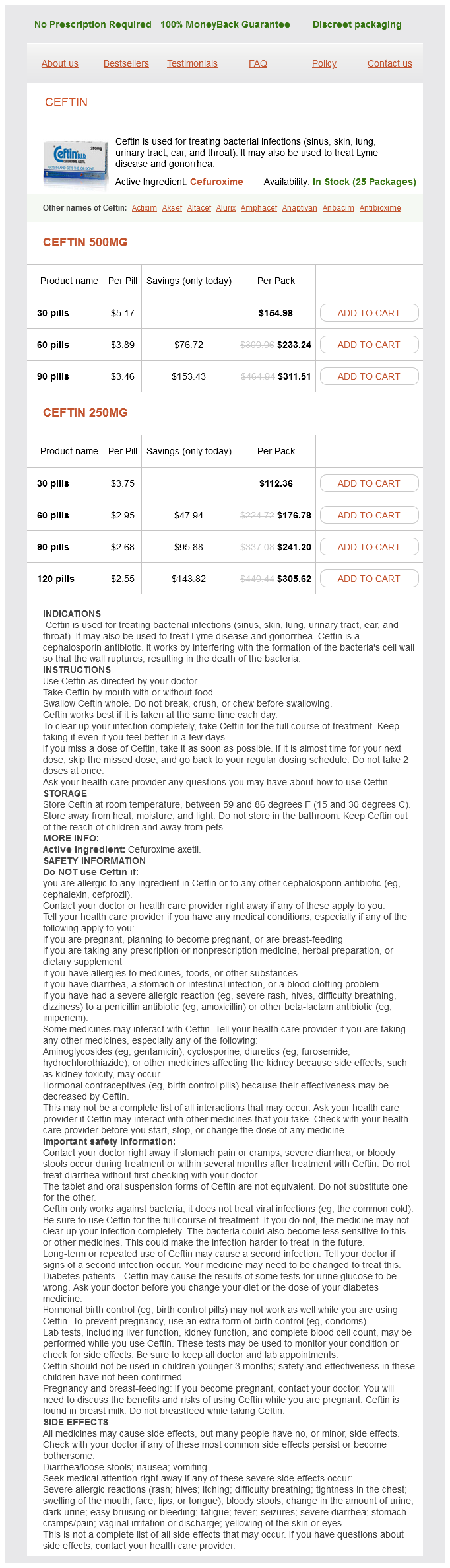
Ceftin, additionally known by its generic name cefuroxime, is a broad-spectrum antibiotic used for treating quite a lot of bacterial infections in different components of the body. It belongs to the cephalosporin family of antibiotics and is on the market in both oral and injectable forms. Ceftin is a generally prescribed medication, identified for its effectiveness in treating infections caused by micro organism.
Urinary tract infections (UTIs) are one other widespread bacterial an infection that Ceftin is used to deal with. UTIs can have an result on different elements of the urinary system, including the kidneys, bladder, and urethra. They are generally brought on by the micro organism Escherichia coli and can trigger symptoms corresponding to pain and burning throughout urination, frequent urge to urinate, and lower stomach pain. Ceftin is effective in treating UTIs, relieving symptoms and preventing issues corresponding to kidney infections.
One of the commonest makes use of of Ceftin is for treating sinus infections. Sinusitis is a common infection of the sinuses, which are cavities positioned within the bones of the face and skull. This infection can cause signs corresponding to a headache, runny nose, facial ache, and issue breathing. Ceftin is effective in treating sinus infections brought on by micro organism, providing aid from symptoms and rushing up the restoration process.
Ceftin is also used for treating pores and skin infections, including cellulitis, impetigo, and folliculitis. These infections can happen when micro organism enter the pores and skin via cuts, wounds, or insect bites. Symptoms of pores and skin infections may embody redness, swelling, and pain. Ceftin works by focusing on these bacteria and clearing the an infection, resulting in improved pores and skin health.
In conclusion, Ceftin is a widely used and effective antibiotic for treating bacterial infections. It is particularly helpful in treating quite a lot of infections in different elements of the body, together with sinuses, skin, lungs, urinary tract, ear, and throat. With its broad-spectrum action and low incidence of unwanted effects, Ceftin is a trusted medicine for combating bacterial infections and promoting better well being. If you're experiencing symptoms of a bacterial an infection, seek the advice of your healthcare supplier to see if Ceftin may be an applicable remedy option for you.
Apart from the frequent infections talked about above, Ceftin is also used for treating Lyme illness and gonorrhea. Lyme disease is a bacterial an infection attributable to a tick chunk and may cause signs such as a rash, fever, and joint pain. Ceftin is used for treating early-stage Lyme illness and can help prevent the spread of the infection. Similarly, Ceftin can also be used for treating gonorrhea, which is a sexually transmitted an infection attributable to the bacteria Neisseria gonorrhoeae.
Ceftin is usually well-tolerated and has a low incidence of unwanted effects. Some frequent side effects embody diarrhea, nausea, and headache. In uncommon circumstances, more severe side effects similar to allergic reactions may occur. It is crucial to inform your doctor when you experience any side effects while taking Ceftin.
In addition to the above, Ceftin can also be used for treating bacterial infections of the ear and throat. These infections may be attributable to streptococcus and Haemophilus influenzae and may end up in signs corresponding to ear ache, sore throat, and difficulty swallowing. Ceftin is an effective treatment for these kind of infections and might present reduction from signs and promote quicker recovery.
In those instances treatment for sinus infection uk buy ceftin master card, aggressive risk factor control with statins and beta blockade should still be implemented. Cardiac arrhythmias may then occur in upward of 6070% of all stroke patients [6,8]. The incidence of serious arrhythmias is highest in first 24 h post admission with up to 25% of patients sustaining a serious arrhythmia in the first 3 days post stroke [10]. Only about 25% of patients have clinical symptoms related to these arrhythmias, however, so continued cardiac telemetry monitoring of all stroke patients while in hospital is essential. Cardioembolic strokes comprise 1430% of all ischemic strokes and atrial fibrillation may represent 50% of these cardioembolic strokes [13]. Serious arrhythmias, however, may only be detected after stroke occurrence and may be directly related to the size or location of stroke. Serious tachyarrhythmia due to atrial fibrillation are particularly frequent in acute ischemic stroke. The extent to which some of these arrhythmias represent a preexisting or otherwise undetected cardiac condition is unknown, but development of arrhythmias should prompt a diagnostic evaluation for underlying cardiac dysfunction. In particular, depolarization abnormalities are of particular concern as they are associated with a greater risk of ventricular tachycardia or fibrillation. The cause of cardiac dysrhythmias likely reflects increased sympathetic tone and alterations in blood pressure and heart rate variability that are common post stroke [6,810]. Many stroke patients who develop cardiac arrhythmias have normal cardiac function, however, and a central nervous system mechanism has been suggested for many of these arrhythmias [6,9,10]. Evidence suggests that right insula control of sinoatrial function is also the basis for an increased risk of bradycardia and hypotension that has been observed with right insular strokes. Conversely, tachycardia and arterial hypertension seem to be more common in patients with left insular lesions, though supraventricular arrhythmias usually occur more frequently in right hemispheric strokes possibly due to loss of parasympathetic control from the right insula [6,9]. Regardless, the evaluation and management of poststroke arrhythmia depends on the type of arrhythmia and whether the patient is asymptomatic or having signs of hemodynamic instability. A neurogenic etiology remains a diagnosis of exclusion and a search for an underlying intrinsic cardiac etiology should be sought including structural cardiac abnormalities. Treatment should focus on controlling the arrhythmia, as necessary, and treating the underlying cause of the arrhythmia. In particular, correction of electrolytes abnormalities with maintenance of normal serum electrolyte levels is essential to prevent acute stroke patients from progressing to a potentially fatal arrhythmia. Three-year follow-up and event rates in the international Reduction of Atherothrombosis for Continued Health Registry. Risk of myocardial infarction and vascular death after transient ischemia attack and ischemic stroke: as systematic review and meta-analysis. Cardiac troponin elevation, cardiovascular morbidity, and outcome after subarachnoid hemorrhage. Cerebrogenic cardiac arrhythmias: cortical lateralization and clinical significance. Although they can present in a myriad of manifestations, patients will usually have a focal onset of neurological symptoms at admission hinting toward this diagnosis. Coronary risk evaluation in patients with transient ischemic attack and ischemic stroke: a scientific statement for healthcare professionals form the stroke council and the council on clinical cardiology of the American Heart Association/American stroke association. There is an independent, graded relationship between the two, and hypertension is therefore aggressively treated in the outpatient environment in order to reduce future stroke risks [1]. In the face of longstanding hypertension, which leads to morphological changes in the vessel walls, this Primer on Cerebrovascular Diseases, Second Edition dx. It is not well established how long the acute stroke period lasts; however depending upon the pathology, this may be anywhere from 24 h to 7 days during which there may be a loss of cerebral autoregulation following the acute stroke. Extreme arterial hypertension is detrimental because it leads to encephalopathy, cardiac complications, and renal insufficiency. Extreme arterial hypotension, on the other hand, is also clearly detrimental by decreasing perfusion to multiple organs, especially the ischemic brain, exacerbating the ischemic injury. This is further associated with an increased tendency for hemorrhagic transformation of an ischemic lesion, especially in the face of pharmacological agents such as antifibrinolytics, anticoagulants, or antiplatelet therapy. Moderate arterial hypertension then, during the acute phase, may be advantageous by improving cerebral perfusion of the ischemic tissue without exacerbating edema or leading to hemorrhagic transformation [6]. There is a U-shaped curve for mortality and morbidity when an acute stroke patient is hypoor hypertensive [7]. This study was terminated prematurely; however, continuation of the antihypertensives did not reduce 2-week mortality or morbidity and was not associated with an increase in adverse events [9]. It is reasonable to discontinue or reduce premorbid antihypertensive regimens at the onset of acute ischemic stroke as swallowing is often impaired, and responses to these medications may be less predictable during the acute phase. It is then reasonable to initiate gradual long-term antihypertensive therapy after the initial 24 h from stroke onset in most patients. The prevalence of hypertension among patients with a recent ischemic stroke is about 70% [14]. Another metaanalysis of 10 randomized trials concluded that treatment with antihypertensive drugs was associated with a significant reduction in recurrent strokes [17]. There was no difference between the target groups with regard to the composite outcome of stroke, myocardial infarction, and vascular death [18]. The optimal drug regimen to achieve the recommended level of reductions is uncertain due to limited direct comparisons; however, available data suggest diuretics or a combination or diuretics with angiotensinconverting enzyme inhibitor may be useful [19]. The modifying influence of prolonged antihypertensive treatment on the tolerance to acute, drug-induced hypotension.
Rare: With buccal form-diarrhea bacteria heterotrophs discount ceftin 500 mg on-line, headache, taste changes, nausea or vomiting, stomach pain. Before you start, consult your doctor if: · You are allergic to anything that touches your skin. What drug does: Destroys fungus cell membrane causing loss of essential elements to sustain fungus cell life. What drug does: Inactivates enzyme and facilitates movement of fluid (aqueous humor) into and out of the eye. Infrequent: Eye symptoms: pain, changes in Continue, but call doctor right vision, blurred vision, discharge or away. Wait at least 15 minutes after putting eye drops in before you put in your soft contact lenses. Before you start, consult your doctor if: · You have eye infection or other eye disease. What drug does: Appears to reduce production of aqueous humor (fluid inside eye), thereby reducing pressure inside eye. Before you start, consult your doctor if: · You have asthma, a bronchial disorder or pulmonary disease. Over age 60: No special problems expected, but visit your doctor on a regular basis while using this drug. Prolonged use: Talk to your doctor about the need for follow-up medical examinations to check pressure inside eye. Potential interactions that may occur are similar to those listed in Possible Interactions With Other Drugs under Beta-Adrenergic Blocking Agents. What drug does: this medicine is a topically applied carbonic anhydrase inhibitor that helps decrease production of aqueous humor (the fluid in the eye) and lowers the pressure inside the eye. Prolonged use: Schedule regular appointments with your eye doctor for eye examinations to be sure the medication is controlling the glaucoma. Others: · If you have any eye infection, injury or wound, consult doctor before using this medicine. Your doctor may switch antiglaucoma drugs for a period of time to return effectiveness. Skin & sunlight: Safety and effectiveness in this age group have not been established. Others: · Advise any doctor, dentist or pharmacist whom you consult that you use this medicine. What drug does: Helps lower intraocular eye pressure by increasing drainage of fluid (aqueous humor) out of the eyeball. Before you start, consult your doctor if: · You plan to have eye or dental surgery. Driving, piloting or hazardous work: Your vision may be blurred or there may be a change in your near or far vision or night vision for a short time after drug use. What drug does: Slows formation of uric acid by inhibiting enzyme (xanthine oxidase) activity. Infrequent: Drowsiness, diarrhea, stomach ache or pain, nausea or vomiting without other symptoms, headache. Prolonged use: Talk to your doctor about the need for follow-up medical examinations or laboratory studies to check liver function, kidney function and serum uricacid levels. Chlorpropamide Chlorthalidone Cyclosporine Diuretics, thiazide* Mercaptopurine Probenecid Theophylline Increased effect of chlorpropamide (with allopurinol). The drug product may contain only the antihistamine or the antihistamine may be an ingredient in a combination drug product that treats multiple allergy/cold symptoms. Antihistamines may be taken daily or only when you have symptoms or as a preventive. What drug does: · Blocks action of histamine after an allergic response triggers histamine release in sensitive cells. Note: Other side effects that occur may be due to other ingredients in the drug product. Adverse reactions and side effects may be more frequent and severe than in younger persons, especially urination difficulty, diminished alertness and other brain and nervous-system symptoms. Skin & sunlight: May cause rash or intensify sunburn in areas exposed to sun or sunlamp. Danger increases if you drink alcohol or take medicine affecting alertness and reflexes, such as other antihistamines, tranquilizers, sedatives, pain medicine, narcotics and mind-altering drugs. Mind-altering drugs* Narcotics* Sedatives* Sleep inducers* Tranquilizers* Increased risk of side effects. What drug does: · Blocks action of histamines which are released in the body during an allergic reaction (such as to seasonal pollens). Histamines cause itching, swollen tissues, sneezing, runny nose and eyes and other symptoms. Rare: · Extreme drowsiness, severe or frequent nosebleeds, rapid or forceful heartbeat, nasal perforation (pain and swelling). Infants & children up to age 18: · Azelastine is approved in children 12 years and older for vasomotor rhinitis and in children 5 years and older for allergic rhinitis. What drug does: Blocks action of histamine after an allergic response triggers histamine release in sensitive cells. Infrequent: Increased appetite, doctor weight gain, mild stomach or intestinal problems, cold or flu-like symptoms. Longer use may be recommended by your doctor depending on the disorder being treated. Skin & sunlight: Rarely, may cause rash or intensify sunburn in areas exposed to sun or ultraviolet light (photosensitivity reaction). Prolonged use: · May lead to tardive dyskinesia (involuntary movement of jaws, lips, tongue, chewing).
Ceftin Dosage and Price
Ceftin 500mg
- 30 pills - $154.98
- 60 pills - $233.24
- 90 pills - $311.51
Ceftin 250mg
- 30 pills - $112.36
- 60 pills - $176.78
- 90 pills - $241.20
- 120 pills - $305.62
It facilitates the early detection of neurovascular disease antibiotic impregnated cement order ceftin 250 mg without a prescription, refines risk assessment, aids in the selection of individualized therapies, and monitors the efficacy of such therapies in a sensitive and quantitative manner. As the era of personalized medicine is on the horizon, molecular imaging and its theranostic application will likely play a pivotal role in both basic research and clinical practice for the improved treatment and prevention of stroke. Will molecular optical imaging have clinically important roles in stroke management, and how Accumulation of ultrasmall superparamagnetic particles of iron oxide in human atherosclerotic plaques can be detected by in vivo magnetic resonance imaging. Direct image of cerebral thromboemboli using computed tomography and fibrin-targeted gold nanoparticles. In vivo imaging of apoptosis in patients with acute stroke: correlation with bloodbrain barrier permeability. Tracking the inflammatory response in stroke in vivo by sensing the enzyme myeloperoxidase. Much of the gas and nutrient exchange occurs at the capillary level, so that understanding blood flow at this scale is important for both normal and disease physiology. Advancements in optical microscopy now enable researchers to assess flow in even the tiniest vessels of the cerebrovasculature. The smallest capillaries are just large enough to let red blood cells squeeze through (about 3 m diameter in mice) so that they can be visualized easily by optical methods. In brain, however, most of the vessels are in the depth of the tissue, so it was not until the development of two-photon microscopy that studies of blood flow in the capillaries became common [3,7]. Since then, several other methods have been developed that can image with sufficient depth and spatial resolution to be useful in studies of capillary structure and function. Two-photon microscopy (also known as multiphoton microscopy, two-photon excited fluorescence microscopy, and two-photon laser scanning microscopy) has become a powerful experimental tool for in vivo studies of not only blood flow, but also many other cellular measurements, and hence it is commonly available. This chapter will describe some of the experimental and measurement techniques used to measure capillary blood flow, focusing on two-photon approaches. For in vivo studies, fluorescent labels can be endogenous or exogenous dyes, but many studies take advantage of the specificity of using genetic strategies to label particular cells or structures. For brain, a window of either glass or merely thinned bone is produced above the region of interest. The easiest way to label the blood vessels is an intravenous injection (usually in rodents via the tail or saphenous vein or retro-orbitally) of dextran-conjugated dyes. The length of time the dye stays in the circulation depends on the size of the dextran; 50100 L of 5% w/v dye conjugated to a dextran of 70 kDa dissolved in saline lasts several hours in mice. Dextran-conjugated dyes are available in many colors, so it is possible to choose a color that does not overlap other indicators in the experiment. In adult brain, normal blood vessels, including individual capillaries, are anatomically stable for many months. Because the pattern of branches is unique, the vessels provide a convenient way of repeatedly finding the same capillaries, neurons, or other cells. One can start with the large vessels, which are easy to see on the brain surface, and follow the branches down to specific capillaries reliably across weeks to months of imaging (analogous to following a city map to find a specific address). Circulating cells, including red and white blood cells, do not take up the dextran-conjugated dye, so they appear as dark patches within the vessels. Previous studies have also used red blood cells that were extracted, labeled in vitro, and reinjected, but this complicated procedure is not necessary with two-photon imaging. Since red blood cells are the vast majority of cells in circulation, most blood flow speed measurements described here rely on the red blood cells. In fact, the occasional white blood cell often temporarily decreases the blood flow speed in a capillary, but no extra label needs to be added to see this effect because these cells also show up as dark regions against the fluorescent dye. Blood cells show up as dark areas when the blood plasma is labeled with intravenous dye injections (red, Texas Red dextran). Red blood cells, nucleated white blood cells, and platelets can be distinguished with injections of rhodamine 6G (green) and Hoechst (blue). Stalled cerebral capillary blood flow in mouse models of essential thrombocythemia and polycythemia vera revealed by in vivo twophoton imaging. Other fluorescent labeling techniques can also be used to label additional types of cells. Transgenic mice may have monocytes or other cells labeled by fluorescent protein expression. In some cases, labeled cells are taken from a donor animal and injected into a recipient in adoptive transfer. Several exogenous indicators also provide convenient ways to label particular cell types. It will not label cells in the healthy brain because it does not cross the bloodbrain barrier, but the nuclei in circulating cells such as white blood cells will fluoresce at blue wavelength. Since platelets do not have nuclei, Hoechst will not label platelets and provides a convenient way to distinguish thrombi in capillaries from leukocyte plugs when using rhodamine 6G [10]. Confocal microscopy works in a similar way and has also been used to image capillaries in the brain, but cannot image as deep as two-photon microscopy because of the effect of light scattering in the tissue. The scanning approach to image formation limits the speed of events that can be captured. The majority of two-photon microscopes scan across the image region one line at time and require at least 0. Most red blood cells in capillaries are moving fast enough that full frame images will not capture the same blood cells in successive frames, making it impossible to quantify blood flow speed. To increase time resolution the laser focus is repeatedly scanned along the length of a vessel segment, called a "line-scan," instead of scanning a full image plane, essentially capturing a very thin image in the middle of the blood vessel. Scan speeds are generally fast enough that they will easily capture the same red blood cells in multiple successive line-scans, thus enabling the speed of the cells to be measured. This data is often displayed by aligning the sequential linescans so that time is along the vertical axis and path is along the vessel on the horizontal axis, also known as a kymograph, or spacetime image. A stationary object, which is always in the same part of the vessel, will result in a straight, vertical line.