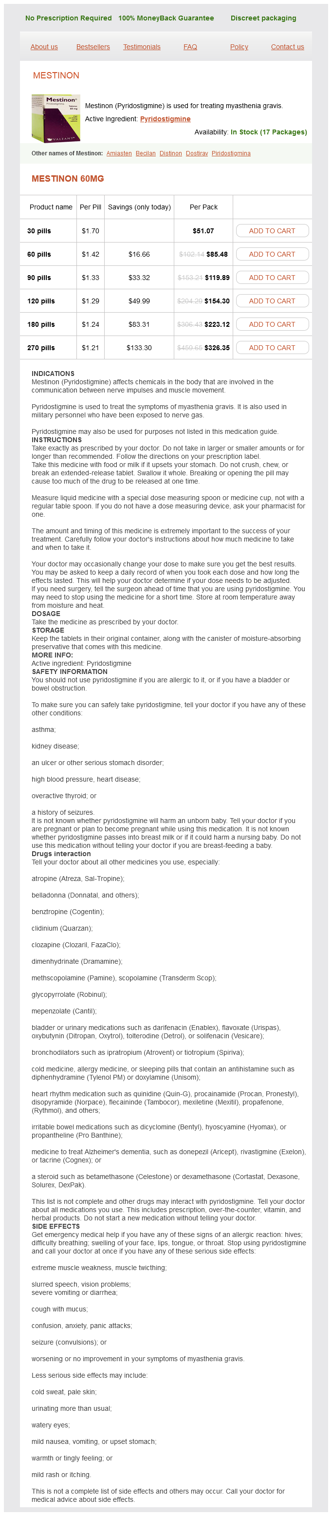
Pyridostigmine Dosage and Price
Mestinon 60mg
- 30 pills - $51.07
- 60 pills - $85.48
- 90 pills - $119.89
- 120 pills - $154.30
- 180 pills - $223.12
- 270 pills - $326.35
There were no significant differences in operative time spasms of the larynx cheap pyridostigmine online amex, operative blood loss, and warm ischemia time, suggesting that robotic assistance enabled those less experienced with laparoscopy to achieve good outcomes. A larger, contemporary single-surgeon study found no difference in the techniques with respect to blood loss and positive surgical margins; however, the robotic approach had advantages of shorter operative and warm ischemia time as well as length of hospitalization (Wang et al, 2009). Since 2013, with robotic partial nephrectomy, it has been feasible to achieve outcomes similar to those obtained with laparoscopic partial nephrectomy (Scoll et al, 2010; Benway et al, 2009). Rates of positive surgical margin are <5%, and complications typically occur in <10% of patients. Potential complications include wound infection, urinary leak/ urinoma, and ileus. Newer techniques have been developed and tested to obtain hemostasis, including the use of hemostatic agents, thermal energy devices, and novel sutures and suturing aids. In addition, selective clamping of renal artery branches or omission/early release of vascular occlusion has been performed to reduce the potential for renal injury. Long-term data are required to confirm cancer control equivalent to radical nephrectomy and/or open partial nephrectomy and preservation of renal function. Most agree, however, that the availability of the robot permits surgeons with less experience with laparoscopy to perform the procedure and shortens the learning curve for the operation and has led to widespread adoption and acceptance (Foerster et al, 2018). In addition, the costs of disposables associated with the robot exceed those of traditional laparoscopy. Bolenz et al (2010) compared the costs associated with robotic, laparoscopic, and open prostatectomy for prostate cancer in 643 patients treated over a 6-year period. Robotic prostatectomy was associated with the highest cost, due to differences in surgical supply and operating room cost. Even with improvements in operative time, rapid time to discharge, and increase in surgical volume, robotic prostatectomy is likely to remain more expensive than other techniques (Lotan, 2010). Given the unproven improvement in outcomes with robotic assistance, physicians should be cautious in the wholesale application to all minimally invasive operations and attempts should be made to systematically study newer surgical technologies. In the future, these economic factors will only be more relevant for both surgeons and patients, given the explosion of healthcare expenditure, greater scrutiny of outcome and quality of care, and inevitable cost-cutting measures in the era of healthcare reform. The advantages may be greater in this domain, given the preponderance of reconstructive, rather than extirpative, procedures. Conversely, the pediatric population poses unique challenges-limited working space within the body, need for smaller ports and instruments, restricted access to the smaller patient by the large surgical cart, and limited haptics on delicate tissue. In addition, the relative benefits of minimally invasive surgery on postoperative recovery and morbidity may be less significant than in the adult. Although a survey of parental satisfaction of open and robotic pyeloplasty using a validated surgery demonstrated greater satisfaction with the robotic approach, the differences were not at great as anticipated (Freilich et al, 2010). The most common robotic operation in children is pyeloplasty, and the early cumulative experience demonstrates that the outcomes are not significantly different from open pyeloplasty (Lee et al, 2006; Olsen et al, 2007; Song et al, 2017). Other applications of the robot include heminephrectomy for nonfunctioning polar moiety in a duplicated system, extravesical ureteral reimplantation, and bladder augmentation. The role of robotics in operations such as adrenalectomy and total nephrectomy are unclear, given the ease in performing the procedure via traditional laparoscopy and inability to take advantage of the added dexterity and precision provided by the robot. Currently, there are no established systems to ensure surgeon competency and safety with robotic urological surgery. These issues have been raised by a panel of experts (Zorn et al, 2009), and future guidelines should be expected to help standardize the process of robotic education, training, proctoring, and credentialing. Tools have been developed to facilitate the ease of surgery and ergonomics, including smaller deflectable-tip laparoscopes, bent and articulating laparoscopic instruments, and single-port access systems. Tan J et al: Robotic-assisted versus open radical cystectomy in bladder cancer: A meta-analysis of four randomized controlled trials. Ureteral obstruction leads to hydroureteronephrosis, and eventually kidney atrophy that may terminate in renal failure. Furthermore, obstruction often is complicated by infection, which can result in systemic complications and cause additional damage to other organs. Etiology Congenital anomalies, more common in the urinary tract than in any other organ system, are generally obstructive. Among the common causes are (1) urethral stricture secondary to infection or injury; (2) benign prostatic hyperplasia or cancer of the prostate; (3) vesical tumor involving the bladder neck or one or both ureteral orifices; (4) local extension of cancer of the prostate or cervix into the base of the bladder, occluding the ureters; (5) compression of the ureters at the pelvic brim by metastatic nodes from cancer of the prostate or cervix; (6) ureteral stone(s); (7) retroperitoneal fibrosis or malignant tumor; and (8) pregnancy. The upper tracts are damaged secondarily by ureterovesical obstruction or reflux and, often, by complicating infection. Severe constipation, especially in children, can cause bilateral hydroureteronephrosis from compression of the lower ureters. Elongation and kinking of the ureter secondary to vesicoureteral reflux commonly lead to ureteropelvic obstruction and hydronephrosis. Unless a voiding cystourethrogram is obtained in children with this lesion, the primary cause may be missed and improper treatment undertaken. Congenital the common sites of congenital narrowing are the external meatus in boys (meatal stenosis) or just inside the external urinary meatus in girls, the distal urethra (stenosis), posterior urethral valves, ectopic ureters, ureteroceles, and the ureterovesical and ureteropelvic junctions (Beganovic et al, 2007; Tan and Smith, 2004). Another congenital cause of urinary stasis is damage to sacral roots 24 as seen in spina bifida and myelomeningocele. Pathogenesis and Pathology Obstruction and neuropathic vesical dysfunction have the same effects on the urinary tract. These changes can best be understood by considering the effects of (1) a severe meatal stricture on the lower tract (distal to the bladder neck), (2) a large obstructing prostate on the midtract (bladder), and (3) an impacted stone in the ureter on the upper tract (ureter and kidney). Lower Tract (eg, Urethral Stricture) Hydrostatic pressure proximal to the obstruction causes dilation of the urethra. If the urine becomes infected, urinary extravasation may occur, and periurethral abscess can result. Acquired Acquired obstructions are numerous and may be primary in the urinary tract or secondary to retroperitoneal lesions 178 B.
Prognosis Survival in testicular cancer has improved dramatically over the past several years spasms heat or ice pyridostigmine 60 mg purchase with visa, reflecting the continuing improvement and refinement in combination chemotherapy. Higher-stage disease treated by orchiectomy and primary chemotherapy has a 5-year disease-free survival rate of 3575%, yet the lower value comes from older series in which more crude chemotherapy regimens were employed. Patients with bulky retroperitoneal or disseminated disease treated with primary chemotherapy followed by surgery have a 5-year disease-free survival rate of 5580%. Treatment and Prognosis Radical orchiectomy is the initial treatment for Leydig cell tumors. Clinical staging is similar to that for germ cell tumors, and levels of the 17-ketosteroids can be helpful in distinguishing between benign and malignant lesions. Because of the rarity of these lesions, the role of chemotherapy remains to be defined. Prognosis is excellent for benign lesions, while it remains poor for patients with disseminated disease. Epidemiology and Pathology Sertoli cell tumors are exceedingly rare, composing <1% of all testicular tumors. A bimodal age distribution is seen: 1-year old or younger and the 20- to 45-year-old age group. Benign lesions are well circumscribed, while malignant lesions show ill-defined borders. Microscopically, tumors appear heterogeneous with mixed amounts of epithelial and stromal components. Sertoli cells are columnar or hexagonal cells with a large nucleus and solitary nucleolus and contain vacuolated cytoplasm. Three types will be considered: Leydig cell tumors, Sertoli cell tumors, and gonadoblastomas. Epidemiology and Pathology Leydig cell tumors are the most common nongerm cell tumors of the testis and account for 13% of all testicular tumors. They follow a bimodal age distribution: the 5- to 9-year-old and the 2535-year-old age groups. The cause of these tumors is unknown; unlike germ cell tumors, there is no association with cryptorchidism. Pathologic examination reveals a small, yellow, wellcircumscribed lesion devoid of hemorrhage or necrosis. Microscopically, hexagonally shaped cells with granular, eosinophilic cytoplasm containing lipid vacuoles are seen. Reinke crystals are fusiform-shaped cytoplasmic inclusions that are pathognomonic for Leydig cells. Virilization is often seen in children, and gynecomastia may be present in 30% of adults. Because of the rarity of these tumors, minimal endocrine data on these patients are available. Most of these tumors occur in patients younger than 30 years, although the age distribution ranges from infancy to >70 years. Gross examination reveals a yellow or gray-white lesion that can vary in size from microscopic to >20 cm and may exhibit calcifications. Microscopically, three cell types are seen: Sertoli cells, interstitial cells, and germ cells. Some reports support adjuvant chemotherapy for primary testicular lymphoma, with improved survival rates of up to 93% after 44 months of follow-up. Leukemic Infiltration of the Testis the testis is a common site of relapse for children with acute lymphocytic leukemia. Bilateral testicular irradiation with 20 Gy and reinstitution of adjuvant chemotherapy constitute the treatment of choice. Clinical Findings the clinical manifestations are predominantly related to the underlying gonadal dysgenesis and are discussed elsewhere in this book. It is noteworthy that four-fifths of patients with gonadoblastomas are phenotypic females. In the presence of gonadal dysgenesis, a contralateral gonadectomy is recommended because the tumor tends to be bilateral in 50% of cases in this setting. The most common primary site is the prostate, followed by the lung, gastrointestinal tract, melanoma, and kidney. The typical pathologic finding is neoplastic cells in the interstitium with relative sparing of the seminiferous tubules. Epidemiology and Pathology Lymphoma is the most common testicular tumor in a patient older than 50 years and is the most common secondary neoplasm of the testis, accounting for 5% of all testicular tumors. It may be seen in three clinical settings: (1) late manifestation of widespread lymphoma, (2) initial presentation of clinically occult disease, and (3) primary extranodal disease. Gross examination reveals a bulging, gray or pink lesion with ill-defined margins. Epidemiology and Pathology Extragonadal germ cell tumors are rare, accounting for approximately 3% of all germ cell tumors. Debate continues over whether these lesions originate from "burned-out" testicular primaries or originate de novo. Most retroperitoneal tumors have their origin from a testicular primary, whereas mediastinal germ cell tumors are truly ectopic.
Lowering the body temperature to less than 25° C spasms after hysterectomy 60 mg pyridostigmine purchase with amex, as is commonly done in cardiovascular surgery, results in a transient but mild thrombocytopenia secondary to platelet sequestration in the spleen and liver. Platelet count and function return to baseline values on return to normal body temperature. In a few cases, severe thrombocytopenia, marked impairment of platelet function, and activation of fibrinolysis and intravascular coagulation may develop. Stored blood contains platelets whose viability is severely impaired by the effects of storage and temperature. Under these conditions, the dead or damaged platelets are rapidly sequestered by the reticuloendothelial system of the patient. This situation is only rarely encountered, however, because the practice of transfusing whole blood has been replaced virtually completely by the use of specific components. Finally, mild thrombocytopenia may be encountered in patients with chronic renal failure, severe iron deficiency, megaloblastic anemia, postcompression sickness, and chronic hypoxia. In reactive thrombocytosis the platelet count is elevated for a limited period and usually does not exceed 800,000/mL, although platelet counts greater than 1 million/mL are occasionally seen. A marked and persistent elevation in the platelet count is a hallmark of myeloproliferative disorders such as polycythemia vera, chronic myelogenous leukemia, and myelofibrosis with myeloid metaplasia (or primary myelofibrosis). In the myeloproliferative disorder known as essential thrombocythemia, platelet counts typically exceed 1 million/mL and may reach levels of several million. In reactive thrombocytosis, platelet production remains responsive to normal regulatory stimuli. Note the increased number of platelets but reasonably normal platelet morphology, characteristic of reactive thrombocytosis. Results of platelet aggregation tests induced by various agents usually reveal normal platelet function in reactive thrombocytosis. Reactive thrombocytosis is not associated with thrombosis, hemorrhage, or abnormal thrombopoietin levels. It seldom produces symptoms per se and disappears when the underlying disorder is brought under control. Guideline for investigation and management of adults and children presenting with a thrombocytosis. Reactive Thrombocytosis Associated With Hemorrhage or Surgery After acute hemorrhage, the platelet count may be low for 2 to 6 days (if no platelet transfusion) but typically rebounds to elevated levels for several days before returning to the prehemorrhage level. A similar pattern of thrombocytopenia and thrombocytosis is seen after major surgical procedures associated with significant blood loss. In both cases the platelet count typically returns to normal 10 to 16 days after blood loss. Postsplenectomy Thrombocytosis Removal of the spleen typically results in platelet counts that can reach or exceed 1 million/mL regardless of the reason for splenectomy. After splenectomy, one would expect an initial increase in the platelet count of approximately 30% to 50%. For unknown reasons, the platelet count, however, far exceeds levels that could result from rebalancing of the circulating platelet pool to incorporate the splenic platelet pool. Unlike after blood loss from hemorrhage or other types of surgery, the platelet count reaches a maximum 1 to 3 weeks after splenectomy and remains elevated for 1 to 3 months. After iron therapy is started, the platelet count usually returns to normal within 7 to 10 days. It is believed that iron plays some role in regulating thrombopoiesis, because treatment of the iron deficiency with iron replacement has resulted in a normalization of the platelet count in thrombocytopenic patients and has been reported to induce thrombocytopenia in patients with normal platelet counts. Thrombocytosis may be found in association with rheumatoid arthritis, rheumatic fever, osteomyelitis, ulcerative colitis, and acute infections. In rheumatoid arthritis the presence of thrombocytosis can be correlated with activation of the inflammatory process. Patients with hemophilia often have platelet counts greater than normal limits, even in the absence of active bleeding. Kawasaki disease is a disorder caused by inflammation of the walls of small and medium-sized arteries throughout the body. It is also known as mucocutaneous lymph node syndrome because it affects lymph nodes, skin, and mucous membranes in the mouth, nose, and throat. The highest incidence of Kawasaki disease is found in Japan and in individuals of Japanese descent, although the disease seems to occur in most, if not all, ethnic groups. It is a self-limited acute vasculitic syndrome of unknown origin, although an infectious etiology has been suspected. Although the disease is self-limiting, there can be lifelong sequelae, including coronary artery thrombosis and aneurysms. The acute febrile stage of the disease lasts 2 weeks or longer, with a fever of 40° C or higher, and is unresponsive to antibiotic therapy. The longer the fever continues, the higher the risk of cardiovascular complications. During this phase, the platelet count usually is elevated, and counts of 2 million/mL have been reported. In addition, acute phase reactants such as C-reactive protein and erythrocyte sedimentation rate are elevated, consistent with an inflammatory state. The higher the platelet count, the higher the risk of cardiovascular complications. After the subacute phase comes the convalescent phase, during which all signs of illness disappear and the acute phase reactants subside to normal. Diagnosis is primarily by excluding other diseases that cause similar signs and symptoms.