General Information about Rumalaya
Although Rumalaya is a natural complement, it's important to take certain precautions earlier than beginning the course. Pregnant and breastfeeding women should seek the guidance of a doctor before using the product. Additionally, individuals with pre-existing medical situations like diabetes, hypertension, and bleeding disorders also needs to seek the assistance of their doctor earlier than starting the supplement.
Rumalaya works by targeting the basis reason for joint ache and stiffness – irritation. Inflammation could be attributable to varied components like age, overuse of joints, and chronic well being conditions like arthritis. The lively elements in Rumalaya assist reduce the production of inflammatory enzymes, thereby lowering irritation and ache. The herbs also stimulate the manufacturing of synovial fluid, which acts as a lubricant for joints, selling better movement and flexibility.
Rumalaya is a herbal complement that has been particularly formulated to promote joint health and alleviate joint pain. It is a novel combination of Ayurvedic herbs which were used for lots of of years to enhance joint function and mobility. Let's dive deeper and understand what makes Rumalaya a go-to solution for sustaining wholesome joints.
Rumalaya is a potent blend of herbs like Boswellia, Guggulu, and Shallaki, which have anti-inflammatory properties. These herbs are known to scale back joint irritation and provide reduction from pain and stiffness. The supplement additionally incorporates Indian Bedellium, which is a natural analgesic, and Rasna, which helps with muscle and joint rest.
Rumalaya is available in pill type and may be simply integrated right into a day by day routine. It is beneficial to take one pill twice a day after meals for a minimum of three months to see desired outcomes. However, it is at all times advisable to consult a healthcare skilled before beginning any new supplement.
Rumalaya has a broad range of benefits in relation to joint health. Its highly effective anti-inflammatory properties make it an efficient solution for reducing joint pain, swelling, and stiffness. It can even assist in improving joint mobility and adaptability, making every day tasks simpler for individuals with joint issues. The supplement can additionally be used to stop age-related joint points by maintaining the well being of cartilage and connective tissues.
Precautions:
How Does Rumalaya Work?
In addition to this, Rumalaya has also shown promising results in managing continual conditions like osteoarthritis, rheumatoid arthritis, and spondylosis. The pure ingredients in Rumalaya don't pose any adverse unwanted facet effects and may safely be used for a protracted period.
In conclusion, Rumalaya is a extremely efficient resolution for maintaining wholesome joints and reducing joint ache and stiffness. Its natural composition, coupled with its potent anti-inflammatory properties, makes it a safe and reliable alternative for people of all ages. Whether you are an athlete trying to improve joint perform or an aging grownup trying to maintain joint well being, Rumalaya is an ideal option to keep your joints healthy and pain-free.
In today's world, the place we are continuously juggling a number of responsibilities and main an lively lifestyle, joint health is usually neglected. Joint pain and stiffness can make even the best duties appear challenging and affect our general well-being. This is the place Rumalaya comes into the picture.
The Basics of Rumalaya:
Benefits of Rumalaya:
Usage and Dosage:
Interestingly medications via g-tube cheap rumalaya 60 pills otc, although most of the antineoplastic antibiotics have antibacterial and antifungal properties, their toxicities preclude their routine use in the treatment of infectious diseases. The drugs have the advantage of greater specificity for tissues responsive to * Compounds and their species of derivation include actinomycin D (dactinomycin, Cosmegen) from Streptomyces parvullus; pentostatin (Nipent) from S. Consequently, they are capable of occupying nuclear or membrane steroid receptors, redirecting the feedback mechanism, and inhibiting the normal or abnormal function of the tissue. The therapeutic advantage lies in their proclivity to inhibit uncontrolled cell proliferation without significant direct cytotoxic effects. Antineoplastic hormones are used initially or as adjunctive therapy in the palliative treatment of malignancies susceptible to the drugs, as noted in Table 21. The compounds are used in the initial and secondary treatment of ovarian carcinoma, testicular tumors, and advanced bladder cancer. It is S phase specific and interferes with the conversion of ribonucleotides to deoxyribonucleotides by selectively blocking the enzyme ribonucleotide reductase. It is also used concomitantly with irradiation therapy in the management of squamous cell carcinoma of the head and neck. They are diverse organic substances provided in small quantities in the diet and are found in a variety of chemical forms and structures. Vitamins have assorted essential biochemical roles in contributing to the maintenance of health and have unique therapeutic places in the treatment of related disorders. Originally, most vitamins were used for the prevention of deficiency syndromes associated with inadequate nutritional intake. For instance, in the presence of a deficiency of a particular nutrient and the subsequent induction of the corresponding condition, adequate replacement of the deficient substance resulted in a cure. And until recently, it was reasonable to conclude that for a person following a balanced diet, no additional vitamin supplementation was necessary. This assumption was valid for normal healthy children, adolescents, and adults in the absence of deficiency (with the exception of pregnancy and lactation). This message has not been heeded in recent years, however, as evidenced by the growing consumption (megadoses) and infatuation with vitamin supplementation in the United States. This was encouraged by the misinformation that if vitamins are important for the maintenance of health, then greater ingestion must necessarily provide better health. This belief has been defended by the equivocal results of clinical and scientific studies interpreted out of context. In particular, such studies have concluded that clinical benefits, such as prevention of aging, can be obtained from the beneficial effects of antioxidants against factors that contribute to oxidative stress. The standards are based on periodic review of the scientific evidence for most healthy persons under normal daily stresses. Almost all of these nutrients are provided by a well-balanced dietary intake plan that includes the four basic food groups. The values are based on normal, healthy, height- and weight-adjusted individuals and have changed dramatically over the last 5 years. In general, vitamin supplementation is warranted in situations where a suspected vitamin deficiency contributes to the condition. In the United States, the relative risk for the development of vitamin deficiencies may be higher in the following individuals: 1. Ranges of daily allowances are determined by age, height, and weight (not shown) within group; in general, requirements increase with age, except with some vitamins. Requirements are usually higher during the first 6 months of lactation than during the second 6 months. Vitamins 331 Vitamin toxicity due to overdose is documented in clinical situations but relies on experimental scientific evidence for an adequate description of such conditions. Vitamins are classified according to their biological inclination for distribution and elimination from the body. The water-soluble vitamins are readily eliminated in the urine and consequently, have a low incidence of toxic effects. In contrast, the fatsoluble vitamins are readily distributed throughout the body, with a propensity for storage in lipid-rich tissue. As a result, they tend to accumulate in physiologic compartments, risking potential cumulative toxicity. This article emphasizes the toxicity and deficiencies associated with vitamins and vitamin supplementation whose adverse reactions have been historically and therapeutically documented. It is important to note that natural diets are rarely deficient in single nutrients, and it is rare for clinical manifestations to develop from single causes. Consequently, the detection and diagnosis of vitamin deficiency, or chronic toxicity, are not without ambiguity. Derivatives of vitamin A include retinol, retinal, retinoic acid, and -carotene (a precursor). These physiologically important factors support proper functioning of the reproductive cycle, visual acuity, somatic growth and differentiation, and visual adaptation to darkness, respectively. Oral or parenteral administration of vitamin A (Aquasol A injection, various tablet and capsule formulations) is indicated for the treatment of conditions associated with, but not exclusive to , vitamin A deficiency states. Deficiency syndromes have been associated with kwashiorkor,* xerophthalmia, keratomalacia, and malabsorption syndromes. It is characterized by anemia, edema, swollen abdomen, skin and hair depigmentation, and hypoalbuminemia. The cornea becomes softened and vulnerable to ulceration and infection, which may lead to blindness. Most symptoms resolve with prompt withdrawal of vitamin supplementation, although increased intracranial pressure requires further palliative measures. As noted earlier, the retinoids are derivatives of retinoic acid that promote epithelial cell differentiation, keratinization, and local inflammation. Therapeutically, the desired effect results in the reduction of the severity and formation of the microcomedones characteristic of acne.
At therapeutic and moderately toxic doses kerafill keratin treatment cheap rumalaya 60 pills with amex, dihydropyridines are well recognized to produce reflex increases in heart rate with an increase in left ventricular stroke volume, leading to an increase in cardiac output. With severe overdoses that result in dramatic Ca2+ channel blockage, all Ca2+ channel antagonists exert a negative inotropic effect with depressed cardiac contraction, conduction blockage, hypotension, and shock. The mechanism of hyperglycemia is likely related to the suppressive effect of the drugs on pancreatic -cell insulin release. As the toxicity produces significant morbidity and mortality, general management revolves around providing supportive care, decreasing drug absorption, and augmenting myocardial function with cardiotonic agents. As described earlier, other cardiotonic drugs may include glucagon, atropine, and catecholamines. Adverse effects reported at therapeutic doses include first-dose hypotension, headache, cough, hyperkalemia, dermatitis, renal dysfunction, and angioedema. Among these, the most potentially toxic agent capable of inducing both arterial and venous vasodilation is nitroprusside. Minoxidil undergoes hepatic biotransformation, producing the active N-O sulfate metabolite. The drug has proved to be efficacious in patients with the most severe and drug-resistant forms of hypertension. Toxicity due to extensions of pharmacological effects of the drug, including hypotension, headache, nausea, flushing, palpitation, dizziness, tachycardia, and angina pectoris; the last occurs as a result of baroreflex-induced stimulation of the sympathetic nervous system; 2. Hydralazine-induced autoimmune reactions, including lupus syndrome, vasculitis, serum sickness, hemolytic anemia, and rapidly progressive glomerulonephritis. Tachycardia is caused by the baroreceptor-mediated increase of the sympathetic tone. Hypertrichosis occurs in all patients who receive minoxidil for an extended period of time and is probably a consequence of K+ channel activation. The most common manifestations of diazoxide intoxication are myocardial ischemia, peripheral and systemic edema, and hyperglycemia. Myocardial ischemia may be precipitated or aggravated by diazoxide, and it results from reflex adrenergic stimulation of the heart and from increased blood flow to nonischemic regions. Hyperglycemia appears to result from its inhibition of the secretion of insulin from pancreatic -cells. Cardiovascular Toxicology 313 the short-term toxic effects of nitroprusside are caused by excessive vasodilation and ensuing hypotension. Toxicity may also result from the conversion of nitroprusside to cyanide and thiocyanate, the mechanisms of which are discussed in Chapter 23. Nitroprusside-induced cyanide poisoning is a result of the development of an anion-gap metabolic acidosis. The management of vasodilator intoxication includes general supportive measures and correction of hypotension and cardiac arrhythmias. Ca2+ channel antagonists and -adrenergic receptor antagonists may be useful in the treatment of myocardial ischemia caused by the vasodilators. Discontinuation of nitroprusside administration, followed by oxygen supplementation, is essential for suspected cyanide toxicity due to nitroprusside. As with cyanide toxicity, sodium nitrite and sodium thiosulfate should be given immediately to enhance the transulfuration of cyanide to thiocyanate. Depressed myocardial contractility frequently manifests as vasodilation and hypotension. Quinidine intoxication causes cinchonism, a symptom complex that includes headache, tinnitus, vertigo, and blurred vision. Disopyramide also has strong anticholinergic activity, which can precipitate glaucoma, constipation, dry mouth, and urinary retention. Coma, respiratory depression, seizure, and ventricular dysrhythmia (torsades de pointes) occur in severe sotalol overdoses. Although the mechanism is not understood, pulmonary fibrosis is a known complication of chronic amiodarone therapy-there is currently no effective treatment for the condition, and it carries a poor prognosis. Although the concept that chemical agents could interfere with cell proliferation was known by about the turn of the 20th century, it was not until the end of World War I that the biological and chemical actions of alkylating agents, such as the nitrogen mustards, were understood. In fact, the adverse reactions and toxicities associated with these substances often limit their usefulness and complicate the course of treatment. Normal, proliferating somatic cells spend the majority of their existence in interphase. The G1 phase lasts from several hours to days and is characterized by synthesis of cell organelles and centriole replication (the G 0 phase represents a resting stage or sub-phase of G1). The G2 * Cancer is a general term referring to the uncontrolled growth of cells or tissue, characterized by the formation of a neoplasm, that is, a mass of cells or tissue (tumor). Cancers are classified according to their cellular origin and their benign or malignant state of histogenic differentiation. In general, benign tumors are designated by attaching the suffix -oma to the cell of origin (a fibroma is a tumor of fibrous origin; adenomas and papillomas are of benign epithelial origin). Malignant tumors are classified according to their germ cell layers and organ of derivation. Malignant tumor nomenclature follows that of benign tumors, with some exceptions: carcinoma (neoplasm of epithelial cell origin); sarcoma (tumor of mesenchymal-connective tissue-and endothelial cell origin); melanoma (melanocyte origin); lymphoma (lymphoid tissue origin); leukemia (hematopoietic cell origin); and tumors arising from the nervous system. The extended part of interphase then continues in the M phase with the stages of cell division- prophase, metaphase, anaphase, and telophase. During this time, the M phase begins with mitosis (nuclear division) and ends with cytokinesis (cytoplasmic) division. Antineoplastic agents suppress the proliferation of neoplasms through interaction at one or more phases of cell replication. An agent may be incorporated as a substrate or may inhibit the normal functioning of an enzyme.
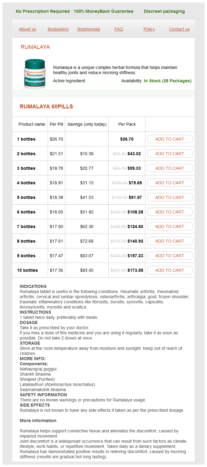
Rumalaya Dosage and Price
Rumalaya 60pills
- 1 bottles - $26.70
- 2 bottles - $43.02
- 3 bottles - $59.33
- 4 bottles - $75.65
- 5 bottles - $91.97
- 6 bottles - $108.28
- 7 bottles - $124.60
- 8 bottles - $140.92
- 9 bottles - $157.23
- 10 bottles - $173.55
As the atrioventricular septum forms during the fifth and sixth weeks medicine 93832 60 pills rumalaya fast delivery, the heart is remodeled to align the developing left atrioventricular canal with the left atrium and ventricle, and the right atrioventricular canal with the right atrium and ventricle. Red arrows indicate the direction of realignment of the atrioventricular canal and outflow tract and formation of the muscular interventricular septum. The blue arrow in C indicates formation of an enlarging slit carved out of the muscular ventricular septum; this is responsible in part for repositioning of the tricuspid orifice to the right, as well as for formation of the moderator band. The frequency of anomalies increases with increasing first-trimester maternal hemoglobin A1c. Hypertrophic cardiomyopathy can develop in response to maternal hyperglycemia in the third trimester. The cranial-lateral wall of the right ventricle has been removed to show the interior of the right ventricular chamber and the presumptive outflow tracts of both ventricles. These swellings are populated by endocardial and neural crest cell -derived cushion cells and develop in a spiraling configuration. They fuse with one another in a cranial-to-caudal direction, forming the conotruncal septum, which separates the aortic and pulmonary outflow tracts. The circular structures to the right of the developing outflow tract illustrate drawings of cross sections at three proximodistal levels. Maternal B-mimetics such as terbutaline can be used to treat a fetus with a critically low heart rate. Maternal infections during pregnancy: Parvovirus can cause fetal myocarditis, with poor ventricular contractility and/or high output cardiac failure due to severe anemia. Congenital rubella is associated with patent ductus arteriosus and pulmonary artery stenosis. Model systems for the study of heart development and disease: cardiac neural crest and conotruncal malformations. Molecular and morphogenetic cardiac embryology: implications for congenital heart disease. The most common cyanotic congenital heart defect seen in infants of diabetic mothers is which of the following Venous return to the fetal heart: Deoxygenated blood from the upper body returns to the heart via the superior vena cava and is directed by the foramen ovale and eustachian valve, across the tricuspid valve and into the right ventricle. Deoxygenated blood from the coronary sinus streams across the tricuspid valve to the right ventricle due to its location adjacent to the tricuspid valve. Deoxygenated blood from the lower body returns via the inferior vena cava and streams across the tricuspid valve. Oxygenated blood from the placenta goes from the umbilical vein primarily into the ductus venosus, with a smaller amount directed to the left portal vein. Ventricular output of the fetal heart: the right ventricle is dominant in fetal life, pumping 55% to 65% of the cardiac output. The right ventricle has greater mass and thus directly affects the filling and ejection of the left ventricle in the fetus. Most of the right ventricular output is directed away from the lungs due to the very high pulmonary vascular resistance in fetal life. Blood is directed across the ductus arteriosus, with one-third delivered to the lower body and two-thirds delivered to the placenta via the umbilical artery for oxygen uptake. Left ventricular output supplies the oxygenated blood from the ascending aorta to the coronary arteries (7% of left ventricular output) and to the brain (55% of the left ventricular output). Prostacyclin is produced by lung distention and is a potent pulmonary vasodilator. Remodeling of the pulmonary vascular bed decreases the muscularity of the proximal arterioles and leads to mature levels of pulmonary vascular resistance by 2 months of age. The flap of the foramen ovale is functionally closed at birth, with the dramatic increase in pulmonary venous return causing left atrial pressure to exceed right atrial pressure. Anatomic closure may not occur for months after birth and remains open in 25% of people. Flow across the ductus arteriosus typically changes from right to left to left to right due to the decrease in pulmonary vascular resistance and increase in systemic vascular resistance after birth. Ductus arteriosus closure is initiated by increased oxygen content and decreased levels of prostaglandins after birth. Ventricular output dramatically increases to meet new increased energy demands due to the work of breathing and thermoregulation: Oxygen consumption triples at birth. The left ventricle becomes the dominant ventricle in the transitional circulation as it pumps at higher pressure and ejects more blood due to the continued patency of the ductus arteriosus. In response to stress, fetal arterial output is redistributed, with increased resistance in the placental and lower body vascular beds to maintain cerebral and cardiac oxygen delivery. When the arterial compensatory mechanisms are exceeded, and fetal heart failure develops, abnormalities are seen in the venous system: the first sign of decompensation is a larger A wave in the inferior vena cava Doppler signal. Development of a reverse A wave in the ductus venosus Doppler trace develops as heart failure progresses. Atrial pulsations in the umbilical venous Doppler trace occur in end-stage fetal heart failure. Hemodynamic Consequences of Perinatal Events Asphyxia impairs the transition of fetal to postnatal circulation: Hypoxia keeps the ductus arteriosus open and promotes pulmonary vasoconstriction. Increased pulmonary vascular resistance promotes tricuspid insufficiency, which increases right atrial pressure and promotes continued right to left shunting at the patent foramen ovale.