General Information about Synthroid
In conclusion, Synthroid is a generally prescribed treatment for the therapy of hypothyroidism and goiters. It is broadly used, safe, and efficient, and has been helping individuals manage thyroid disorders for decades. With correct use and monitoring, Synthroid might help individuals lead a more healthy and extra productive life. However, it is essential to consult a doctor earlier than beginning or making any modifications to the medication.
Another benefit of Synthroid is that it's comparatively easy to use and has a minimal risk of unwanted side effects. Unlike other thyroid medications which will comprise animal-derived hormones, Synthroid is synthetic and has a constant potency. It can also be well-tolerated by most individuals, with only some experiencing mild side effects corresponding to complications, nausea, or hair loss. In rare circumstances, some individuals might have an allergic response or experience a fast heartbeat, irregular heartbeat, or chest pain. It is essential to seek the guidance of a physician if any of these unwanted aspect effects occur.
Synthroid works by replacing the lacking thyroxine within the physique and restoring normal thyroid hormone ranges. It is on the market in pill form in numerous strengths, which permits for individualized therapy primarily based on the severity of the hypothyroidism. The medication is usually taken once a day, preferably on an empty stomach, no less than half-hour earlier than a meal.
Synthroid, also known by its generic name levothyroxine, is a medication used for the treatment of low thyroid hormone levels (hypothyroidism) and sure kinds of goiters. It is a synthetic type of the hormone thyroxine, which is produced by the thyroid gland.
The thyroid gland plays an important position in regulating various bodily capabilities corresponding to metabolism, coronary heart fee, physique temperature, and progress. When the thyroid gland does not produce sufficient thyroxine, it can result in hypothyroidism. This situation may cause symptoms corresponding to fatigue, weight achieve, cold intolerance, hair loss, and depression. If left untreated, it might possibly additionally lead to critical well being problems, together with coronary heart illness, nerve damage, and infertility.
It is essential to take Synthroid exactly as prescribed by a physician. The dosage could additionally be adjusted based on periodic blood checks to guarantee that the treatment is working effectively. It is essential not to miss any doses, as this could disrupt the steadiness of thyroid hormones in the body. Remember to inform the physician of another medications or supplements being taken, as they may intervene with the absorption or effectiveness of Synthroid.
One of the advantages of using Synthroid is its lengthy historical past of protected and efficient use. It has been on the market since 1955 and has been prescribed to tens of millions of people worldwide. It can be permitted by the Food and Drug Administration (FDA) for the treatment of hypothyroidism. This gives sufferers and healthcare suppliers confidence in its reliability and efficacy.
In some instances, Synthroid will not be suitable for people with sure medical conditions, corresponding to heart illness, diabetes, or adrenal gland problems. Pregnant or breastfeeding girls should also seek the assistance of a health care provider earlier than taking Synthroid, as it could possibly have an result on the development of the unborn child or milk manufacturing.
In addition to treating hypothyroidism, Synthroid is also used to shrink an enlarged thyroid gland, also referred to as a goiter. A goiter may be attributable to various factors, together with an iodine deficiency, thyroid nodules, or irritation of the thyroid gland. Synthroid may help reduce the scale of the goiter by regulating thyroid hormone ranges and preventing further enlargement.
By what mechanism can heparin-induced thrombocytopenia actually inaease dot formation With platelet counts of less than about 5000/µL medicine 968 purchase cheap synthroid online, pinpoint hemorrhages (petechlae) may spontaneously occur in the skin or mucous membranes. These are self-limited because the plasma coagulation factors are still intact, and only a small number ofaggregated platelets are needed to provide adequate phospholipids for clotting. The relationship between the likelihood of bleeding and the platelet count is not linear. The bleeding time, a test used clinically to evaluate platelet function, does not even begin to be abnormally prolonged until the platelet count is less than 90,000/µL. Spontaneous bleeding is unlikely until platelet counts are less than 20,000/µL but is still uncommon until counts are less than about 5000/µL. For example, aspirin inhibits platelet aggregation and increases the likelihood of bleeding. When bleeding from thrombocytopenia does occur, it is most often mucosal or superficial in the skin. This may create a very confusing picture, because the heparin may have been given therapeutically for another thrombosis; it may be difficult to determine whether the new thrombosis is an extension of the initial clot or a new one resulting from the heparin exposure. When heparin-induced thrombocytopenia and thrombosis do occur, the clinical manifestation of the new thrombosis will depend on the site of the thrombus. Most studies of this disorder suggest that when thrombosis occurs, it is at the site of previous vascular injury or abnormality. Thus, in patients with atherosclerotic vascular disease, arterial thromboses are much more common than venous clots. Patients experience a rapid onset of severe pain, usually in an extremity, with a cool, pale limb. Inherited Hypercoagulable States Etiology the formation of blood clots in otherwise normal vessels is distinctly abnormal because the coagulation system in mammalian species is both positively and negatively balanced by so many factors. Nonetheless, there are a number of diseases that result in abnormal clotting (thrombod·). Abnormal clotting states may be either primary, in that the abnormalities result from genetic p. Except for hyperprothrombinemia, all lead only to moderate (50%) decreases in the levels of the relevant factors. Despite the relatively modest fall, affected individuals are predisposed to abnormal thrombosis. These disorders are relatively rare in the general population, but they do account for a significant percentage of young patients who come to medical attention with thromboses. Hyperhomocystinemia, an inborn error of metabolism, is also an inherited hypercoagulable state, but because it does not involve the coagulation cascade, it is not further discussed here. Factor Va thus makes an excellent negative control point, so that once clot formation has begun. This amino acid change alters the three-dimensional conformation of the cleavage site within factor Ve. Therefore, loss of this cofactor leads to decreased anticoagulant activity and contributes to the hypercoagulable state. The families that are thrombosis prone are thought to carry additional genetic factors, in addition to protein C deficiency, that increase their risk for thrombosis. Normally, some of the thrombin generated binds to an endothelial cell protein, thro. Protein C deficiency is not all one disease, however, unlike the factor V Leiden abnormality. Type Il deficiency denotes cases with normal protein C levels but low protein C activity. Protein S Deficiency-Protein S deficiency is also an uncommon heterogeneous disorder. Type I protein S deficiency refers to cases with low free and total protein S levels. Protein S, not a protease itself, exposes this site so that protein C can cleave lne. The action of each of the negative control factors-protein S, protein C, and antithrombln-ls shown In color. Because protein S is so crucial, deficiency of protein S also leads to the unregulated procoagulant action of factor Xa. Presumably, this leads to excess thrombin generation when the prothrombinase complex is activated. This is probably the second most common hereditary hypercoagulable state after factor V Leiden. It is the first hereditary thrombophilia associated with overproduction of procoagulant factors. Pathology the pathologic features of thrombi in hypercoagulable states are indistinguishable from those of genetically normal individuals on a gross anatomic or microscopic basis, except that there is a greater likelihood in hypercoagulable states of having a clot in unusual sites (see Clinical Manifestations section). Most of the pathologic features of the hereditary hypercoagulable states consist of laboratory abnormalities. In the evaluation of patients suspected of having a hereditary hypercoagulable state, there are two basic types oflaboratory abnormalities. Prothrombin levels can also be Clinical Manifestations Most thromboembolic events encountered in clinical practice are secondary, not primary. Patients have blood clots usually in the deep veins of the legs for two reasons: (1) because of sluggish blood flow (in high-capacity, low-flow veins) compared with other sites, particularly when inactive (eg, bedridden after surgery or as a result of illness); and (2) because the extremities are more likely to sustain injury than the trunk.
Leukocytoclastic vasculitis lesions are raised and papular because lesional skin is altered and expanded by an intense vasocentric infiltrate containing numerous neutrophils treatment zit best purchase for synthroid. The lesions are purpuric or erythematous because of the extravasated red blood cells that accumulate in the dermis. It would be important to inquire about these symptoms and order laboratory tests to assess liver or renal involvement. The anterior lower legs are the most common locations for such (node-like) lesions to develop. The fact that the patient herself had symptoms of pharyngitis, which were alleviated with antibiotics, is helpful. However, because the antibiotics course was much shorter than required (2 days vs. Once the infection has been eradicated, the skin lesions should subside within several weeks. The history of hiking in a heavily wooded area 2 days before onset of the rash is a helpful clue. However, the finding on physical examination of blisters arranged in straight lines helps make the diagnosis. In this case, poison ivy leaves traced a line across the skin as the patient walked through the brush, and she developed an allergic contact dermatitis in the pattern of the exposure to its sticky, long-lasting oil (known as urushiol). A common misconception regarding Rhus dermatitis is that blister fluid from broken blisters (or even touching the blistered area) causes the eruption to spread. In fact, once the eruption has developed, the urushiol allergen has been irreversibly bound to other proteins or has been so degraded that it cannot be transferred to other sites. In this case, the patient developed large blisters or bullae in response to the contactant at the original sites of contact, the legs. Intense inflammation such as this can result in the autosensitization phenomenon, C. Erythema nodosum is thought to represent a systemic, delayed-type hypersensitivity reaction that localizes to the subcutis for unknown reasons. In erythema nodosum, the inflammatory response consists of lymphocytes, histiocytes, neutrophils, and eosinophils scattered throughout the septal compartment of the subcutis with frequent multinucleated histiocytes. The septa are thickened and may become fibrotic, depending on the density of the infiltrate and the duration of the reaction. Microscopically, evidence of fat necrosis may be seen in the form of an infiltrate of "foamy" (lipid-laden) macrophages at the periphery of subcutaneous lobules or in the form of small stellate clefts within multinucleate macrophages, indicating an element of lipomembranous fat necrosis. She should also be advised to use nongreasy cosmetics, usually those labeled as "noncomedogenic," as well as hair care products without petrolatum. Bacterial factors and sebum breakdown products attract neutrophils to the follicle, thus forming a pustule. Follicular rupture induces an intense inflammatory response in the dermis seen clinically as an inflammatory papule or pustule. Follicular plugging may be corrected with retinoids (vitamin A analogues) either topically or, if the condition is severe enough, orally. Oral antibiotics such as erythromycin or tetracycline are frequently used in addition to topical antibiotics. These agents are not merely antibacterial but are known to have anti-inflammatory properties independent of their antibacterial action. Last, sebum production may be decreased through the use of retinoids, again topically or orally, although oral therapy is much more effective for this purpose, or with antiandrogen medications such as spironolactone and oral contraceptives. Because sarcoidosis is a diagnosis of exclusion, a thorough workup for specific causes is warranted. A skin biopsy should demonstrate changes typical of sarcoidosis (non-caseating granulomata), with negative histochemical stains for mycobacterial and fungal organisms. A chest x-ray film is helpful to rule out tuberculosis and to search for hilar adenopathy. This patient has sarcoidal papules around the edges of the nostrils, a finding known as lupus pernio or nasal rim sarcoidosis. This finding indicates that the patient is at high risk for significant involvement of the tracheobronchial tree or lung parenchyma. Regardless of symptoms and dermatologic presentation, the possibility of pulmonary involvement should always be investigated in all cases of sarcoidosis because it is quite common and sometimes asymptomatic. Sarcoidosis is a nodular dermatitis with histiocytic granulomas situated within the dermis. Sarcoidosis is seen clinically as an elevation (papule, plaque, or nodule) caused by the expansion of the dermis by the infiltrate. Urticaria affects approximately 15-25% of the population, including people of all ages. In some patients, a specific cause can be identified, such as sunlight, water, medication, pressure, vibration, heat, cold, exercise, or emotional stress. Urticaria is the result of mast cell degranulation resulting in a release of histamine and other pro-inflammatory cytokines such as prostaglandins, leukotrienes, and platelet activating factor. While the type I hypersensitivity reaction mediated by IgE is the classic cause for mast cell degranulation, there are in fact many other mediators of mast cell degranulation, including complement activation, physical stimuli (such as exercise or cold temperatures), viral infections, and autoantibodies. The release of histamine causes capillary vasodilation in the superficial dermis with the subsequent extravasation of protein-rich fluid into the superficial aspects of the skin and the development of the urticarial papules and/or plaques.
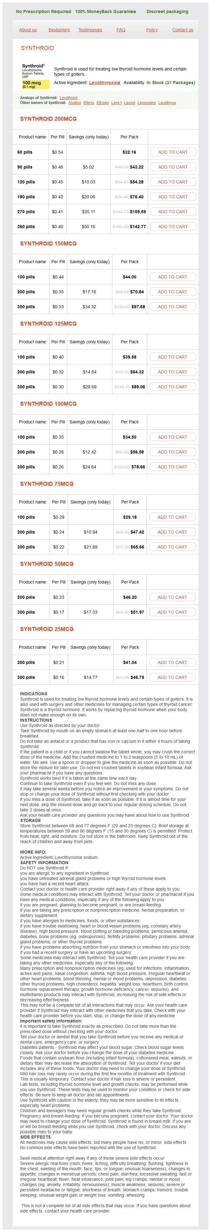
Synthroid Dosage and Price
Synthroid 200mcg
- 60 pills - $32.16
- 90 pills - $43.22
- 120 pills - $54.28
- 180 pills - $76.40
- 270 pills - $109.59
- 360 pills - $142.77
Synthroid 150mcg
- 100 pills - $44.00
- 200 pills - $70.84
- 300 pills - $97.68
Synthroid 125mcg
- 100 pills - $39.58
- 200 pills - $64.32
- 300 pills - $89.06
Synthroid 100mcg
- 100 pills - $34.50
- 200 pills - $56.58
- 300 pills - $78.66
Synthroid 75mcg
- 100 pills - $29.18
- 200 pills - $47.42
- 300 pills - $65.66
Synthroid 50mcg
- 200 pills - $46.20
- 300 pills - $51.97
Synthroid 25mcg
- 200 pills - $41.04
- 300 pills - $46.79
Upon ligand binding symptoms questions order synthroid without prescription, the receptor attaches to enhancer sites in target genes and directly regulates their transcription. The most essential action ofl,25-(0H)p is to stimulate the active intestinal transport of calcium in the duodenum. Calcium also can be absorbed passively through a paracellular route throughout the small intestine. However, particularly at low calcium intakes, the majority of gastrointestinal calcium absorption is mediated by the active vitamin D-mediated process. However, the defect in mineralization results mainly from a decrease in the delivery of calcium and phosphate to sites of mineralization. The net absorption of calcium falls sharply, causing a transient decrease in the serum calcium level. Over the long term, these compensatory mechanisms will result in depletion of skeletal calcium, increased bone resorption, and compromised skeletal integrity. As a result, lntestlnal and renal phosphate reabsorptlon rises to restore serum phosphate back to nonnal. Under normal physiologic: conditions, when phosphate levels rise (eg, high-phosphate diet. Where does the flnal step In the activation of vttamln D take place, and how Is It regulated C cells are neuroendocrlne cells derived from the ultimobranchial body, a structure that fuses with the thyroid. C cells are small, spindle-shaped or polygonal cells distributed throughout the thyroid. They may be present as single cells or arranged in nests, cords, and sheets within the thyroid parenchyma. They are often found within thyroid follicles, are larger than follicular cells, and stain positively for calcltonin. In the kidney, receptors for calcitonin arc localized in the cortical a1cending limb of the Henle loop, whereas in bone, caldtonin recepton arc found on omodasts. The main function of calcitonin is to lower scrum calcium, and this hormone is rapidly released in response to hypercalcemia. Caldtonin inhibits omoclastic bone resorption and rapidly blocks the release of calcium and phosphate from bone. The latter effect is apparent within minutel after the administration of calcitonin. With continued administration of calcitonin, an ·escape· from its effects on serum calcium occurs. The overall importance of calcitonin in the maintenance of calcium homcostasis is unclear. Scrum calcium concentrations arc normal in patients after thyroidcctomy, which removes all functioning C cclla. Similarly, calcitonin typically rises to very high levels in patients with mcdullary carcinoma of the thyroid with no apparent effect on serum calcium levels. Differential procetsing of the caldtonin gene can lead to the production of either calcitonin in C cells or calcitonin gene-related peptide in neurons. Although both calcitonin and calcitonin gene-related peptide have demonstrated clinical effects in pharmacologic doses, the function of the peptides at normal physiologic levels is unknown. Subttantial changes in serum calcium are normally required to modulate the release of calcitonin. Calcitonin secretion in vivo is assessed by measuring serum levels with a two-site radioimmunoa. The prevalence ofhyperparathyroidism is approximately 1:1000 in the United States, and the incidence of the disease increases with age. Chief c:e1l adenomas are the most common cause, accounting for ahnost 85% of all cases. Parathyroid hyperplasia refers to an enlargement or abnormality of all four glands. Distinguishing between hyperplasia and multiple adenomas is challenging and usually requires the examination of all four glands. Key characteristics for judging whether a gland is normal or not are its size, weight, and histologic features. Parathyroid hyperplasia may be part of the autosomal dominant multiple endocrine neoplaaia (. When their glands are examined microscopically, there are usually abnormalities in all four glands. Recurrent hyperparathyroidism, even after initially successful surgery, is common in these patients. The hyperparathyroidism-jaw tumor syndrome and familial isolated hyperparathyroidism are other causes of autosomal dominant hyperparathyroidism. Parathyroid cardnoma is a rare malignancy, but the diagnosis should be considered in a patient with severe hypercalcemia and a palpable cervical mass. At surgery, cancers are firmer than adenomas and more likely to be attached to adjacent structures. It is sometimes difficult to distinguish parathyroid carcinomas from adenomas on hi. Secondary hyperparathyroidism in patients with normal kidney function may be observed in patients with severe calcium and vitamin D deficiency states (see below). This qualitative regulatory defect is more common than truly autonomous secretion. How these two defects interact in the pathogenesis of the disease remains to be fully elucidated. The genetic defects responsible for primary hyperparathyroidism have received considerable attention.