General Information about Urispas
Urispas, additionally identified by its generic name flavoxate, is a medication used to deal with urinary problems in people with sure medical situations. It belongs to a category of medicine called urinary antispasmodics and works by relaxing the muscular tissues within the bladder, thereby decreasing pain, frequency, and urgency of urination.
As with any medicine, Urispas might trigger side effects in some individuals. Common unwanted effects embrace dry mouth, nausea, constipation, and dizziness. If these unwanted effects turn out to be extreme or persistent, it is important to seek the assistance of with a healthcare supplier.
It can be necessary to tell your healthcare supplier of another medications you're taking, as Urispas can interact with sure drugs, similar to antihistamines and antidepressants. It is also not beneficial to drink alcohol whereas taking Urispas.
Another frequent use of Urispas is for individuals with urinary tract infections (UTIs). UTIs are caused by bacteria coming into the urinary tract and may cause painful urination, frequent urination, and a strong urge to urinate. Urispas may help alleviate these signs and also help forestall recurrent UTIs.
Urispas is generally well-tolerated and might present relief for people affected by urinary issues. However, it isn't beneficial for use in people with certain medical situations, similar to glaucoma, an enlarged prostate, or an obstructive gastrointestinal disorder. It is necessary to debate your medical historical past together with your healthcare provider before beginning Urispas.
In conclusion, Urispas is a drugs that may provide reduction from the ache, frequency, and urgency of urination in individuals with sure medical conditions. It is essential to consult with a healthcare supplier earlier than beginning remedy and to follow the prescribed dosage to attain most advantages. With correct use, Urispas can considerably enhance the quality of life for these affected by urinary issues.
One of the primary makes use of of Urispas is for people that suffer from urinary incontinence, which is the involuntary leakage of urine. This situation may be attributable to a wide range of elements, together with bladder muscle spasms, overactive bladder, and nerve injury. Urispas may help management these symptoms and enhance the individual's high quality of life.
In addition, Urispas can be utilized for people with bladder problems, similar to interstitial cystitis and bladder ache syndrome. These situations are characterised by bladder pain and discomfort, and Urispas might help cut back these symptoms by enjoyable the muscle tissue in the bladder.
Urispas is often taken orally, with or with out meals, and the dosage is typically based on the individual's age, medical condition, and response to therapy. It is important to observe the prescribed dosage and to proceed taking the medicine even if signs improve, as stopping the medication abruptly could cause a return of symptoms.
Vitamin K Antagonists Coumarin anticoagulants owe their action to the ability to antagonize the cofactor functions of vitamin K muscle relaxant injections urispas 200 mg buy otc. The -carboxyglutamyl residues bind calcium ions, which are essential for interaction between the coagulation factors and platelet membranes. The reduced vitamin K cofactor is converted to vitamin K epoxide during the reaction. Vitamin K is regenerated from the epoxide by vitamin K epoxide reductase, the enzyme that is inhibited by warfarin. Warfarin treatment results in the production of clotting factors with diminished activity (10% to 40% of normal), due to the lack of sufficient -carboxyglutamyl side chains. Unlike heparin, the anticoagulant effects of warfarin are not observed immediately after drug administration. However, reversal following administration of vitamin K takes approximately 24 hours (the time necessary for degradation of already synthesized clotting factors). It is also used for prevention of venous thromboembolism following orthopedic surgery. Pharmacokinetics Warfarin is rapidly absorbed after oral administration (100% bioavailability with little individual patient variation). However, drugs that have a greater affinity for the albumin-binding site, such as sulfonamides, can displace the anticoagulant and lead to a transient, elevated activity. The mean half-life of warfarin is approximately 40 hours, but this value is highly variable among individuals. After conjugation to glucuronic acid, the inactive metabolites are excreted in urine and feces. Warfarin has numerous drug interactions that may potentiate or attenuate its anticoagulant effect. Minor bleeding may be treated by withdrawal of the drug or administration of oral vitamin K, but severe bleeding may require greater doses of vitamin K given intravenously. Purple toe syndrome, a rare, painful, blue-tinged discoloration of the toe caused by cholesterol emboli from plaques, has also been observed with warfarin therapy. Therapeutic use Dabigatran is approved for the prevention of stroke and systemic embolism in patients with nonvalvular atrial fibrillation. The drug is contraindicated in patients with mechanical prosthetic heart valves and is not recommended in patients with bioprosthetic heart valves. Dabigatran should be used with caution in renal impairment or in patients over the age of 75, as the risk of bleeding is higher in these groups. About one-third of the drug is excreted unchanged in the urine, and the inactive metabolites are excreted in the urine and feces. Edoxaban and betrixaban are minimally metabolized and are eliminated primarily unchanged in the urine and feces, respectively. All of these drugs are substrates of P-gp, and dosages should be reduced (in some cases concomitant use should be avoided) with P-gp inhibitors such as clarithromycin, verapamil, and amiodarone. Currently there is no antidote, but recombinant factor Xa products are in development. Declining kidney function can prolong the effect of these drugs and, therefore, increase the risk of bleeding. Thrombolytic Drugs Acute thromboembolic disease in selected patients may be treated by the administration of drugs that activate the conversion of plasminogen to plasmin, a serine protease that hydrolyzes fibrin and, thus, dissolves clots. Unfortunately, increased local thrombi may occur as the clot dissolves, leading to enhanced platelet aggregation and thrombosis. Strategies to prevent this include administration of antiplatelet drugs, such as aspirin, or antithrombotics such as heparin. Adverse effects Thrombolytic agents do not distinguish between the fibrin of an unwanted thrombus and the fibrin of a beneficial hemostatic plug. These drugs are contraindicated in pregnancy and in patients with healing wounds, a history of cerebrovascular accident, brain tumor, head trauma, intracranial bleeding, and metastatic cancer. Alteplase has a very short half-life (5 to 30 minutes), and therefore, a portion of the total dose is injected intravenously as a bolus, and the remaining drug is administered over 1 to 3 hours, depending on the indication. Tenecteplase has a longer half-life and, therefore, may be administered as an intravenous bolus. Drugs Used to Treat Bleeding Bleeding problems may have their origin in naturally occurring pathologic conditions, such as hemophilia, or as a result of fibrinolytic states that may arise after surgery. The positively charged protamine interacts with the negatively charged heparin, forming a stable complex without anticoagulant activity. Vitamin K Vitamin K1 (phytonadione) administration can stop bleeding problems due to warfarin by increasing the supply of active vitamin K1, thereby inhibiting the effect of warfarin. Because it reverses the effect of dabigatran, thrombosis is the most serious adverse effect of idarucizumab. If a patient is not compliant, then the antiplatelet activity of ticagrelor stops when the drug is missed (since the platelets are not irreversibly inhibited as they would be with aspirin, clopidogrel, or prasugrel). Which anticoagulant for atrial fibrillation avoids the need for renal dose adjustment in this patient All of the other agents are renally cleared to some extent and require dosage adjustments in renal dysfunction. He is diagnosed with a urinary tract infection and is prescribed sulfamethoxazole/trimethoprim. Decreased anticoagulant effect of warfarin Increased anticoagulant effect of warfarin Activation of platelet activity No change in anticoagulation status Correct answer = B. Sulfamethoxazole/trimethoprim has a significant drug interaction with warfarin, such that it inhibits warfarin metabolism. Protamine Vitamin K Idarucizumab A reversal agent does not exist for this medication Correct answer = C.
Circumvallate Papillae Circumvallate papillae are larger than the fungiform or filiform papillae spasms near belly button buy urispas paypal. About 8 to 12 circumvallate papillae are located in the posterior region of the tongue in 507 humans. Numerous excretory ducts from underlying serous (von Ebner) glands located in the connective tissue of the tongue empty their serous secretions into the base of these furrows. Numerous taste buds are located in the stratified epithelium on the lateral sides of each papilla. Foliate Papillae Foliate papillae are well developed in some animals but are rudimentary or poorly developed in humans. Taste Buds Located in the confines of the stratified epithelium of the foliate and fungiform papillae, and on the lateral sides of the circumvallate papillae, are barrel-shaped taste buds. In addition to the tongue, taste buds are found in the epithelium of the soft palate, pharynx, and epiglottis. The epithelial surface of each taste bud contains an opening called the taste pore. Each taste bud occupies the full thickness of the epithelium and contains three main cell types. Located within each taste bud are elongated gustatory (neuroepithelial or taste) cells that extend from the base of the taste bud to the taste pore. The apices of each taste cell exhibit numerous microvilli that protrude through the taste pore. Also present in the taste buds are elongated, supporting sustentacular cells that are less numerous and not sensory. At the base of each taste bud are the basal cells that are undifferentiated and serve as stem cells for the other two cell types. Lymphoid Aggregations: Pharyngeal, and Lingual) Tonsils (Palatine, the tonsils are aggregates of diffuse lymphoid tissue and lymphoid nodules that are located in the oral pharynx. The palatine tonsils are located on the lateral walls of the oral part of the pharynx. These tonsils are lined with stratified squamous nonkeratinized epithelium and exhibit numerous crypts. The pharyngeal tonsil is a single structure situated in the superior and posterior portions of the pharynx. The lingual tonsils are located on the dorsal surface of the posterior third of the tongue and are visible as numerous small bulges composed of massed lymphoid aggregations. The lingual tonsils are lined with a stratified squamous nonkeratinized epithelium. Each tonsil is invaginated by the covering epithelium to form crypts, around which are found lymphatic nodules with germinal centers. The epidermis (11) exhibits stratified squamous keratinized epithelium with desquamating surface cells (10). Beneath the epidermis (11) is the dermis (14) with sebaceous glands (2, 12) associated with hair follicles (4, 15) and the simple tubular sweat glands (16) deeper in the dermis (14). Also visible in the periphery are blood vessels, an artery (6a), and a venule (6b). The transition zone (1) between the skin epidermis (11) and the oral epithelium is a mucocutaneous junction. The internal or oral surface of the lip is 509 lined with a moist, stratified, squamous nonkeratinized oral epithelium (8) that is thicker than the epithelium of the epidermis (11). The surface cells of the oral epithelium (8), without cornification, are sloughed off (desquamated) into the fluids of the mouth (10). Deeper connective tissue contains tubuloacinar and mucus-secreting labial glands (9, 18). The connective tissue of the lip also contains numerous adipose cells (7), blood vessels (6), and numerous capillaries. Because the blood vessels (6) are close to the surface, the color of the blood shows through the overlying thin epithelium, giving the lips a characteristic red color. The oral cavity is lined with a protective mucosa (5) that consists of an outer epithelial layer (epithelium) (5a) and an underlying connective tissue layer, the lamina propria (5b). The dorsal surface of the tongue is rough due to numerous mucosal projections called papillae (1, 2, 6). The slender, cone-shaped filiform papillae (2, 6) are the most numerous and cover the entire dorsal surface of the tongue. Less numerous are the fungiform papillae (1) with a broad, round surface of noncornified epithelium and a prominent core of lamina propria (5b). The core of the tongue consists of crisscrossing bundles of skeletal muscle (3, 7). As a result, the skeletal muscles of the tongue are typically seen in longitudinal, transverse, or oblique planes of section. In the connective tissue (9) around the muscle bundles are arteries (4a, 8a), veins (4b, 8b), and nerve fibers (11). In the lower half of the tongue and surrounded by skeletal muscle fibers (3, 7) is a section of the anterior lingual gland (10). This gland is of a mixed type and contains both mucous acini (10b) and serous acini (10c), as well as mixed acini. The interlobular ducts (10a) from the anterior lingual gland (10) pass into the larger excretory duct of the lingual gland (12) that opens into the oral cavity on the ventral surface of the tongue. The lingual epithelium (2) that covers the circumvallate papilla is stratified squamous epithelium (1). The underlying connective tissue, the lamina propria (3), exhibits numerous secondary papillae (7) that project into the overlying epithelium (1, 2) of the papilla.
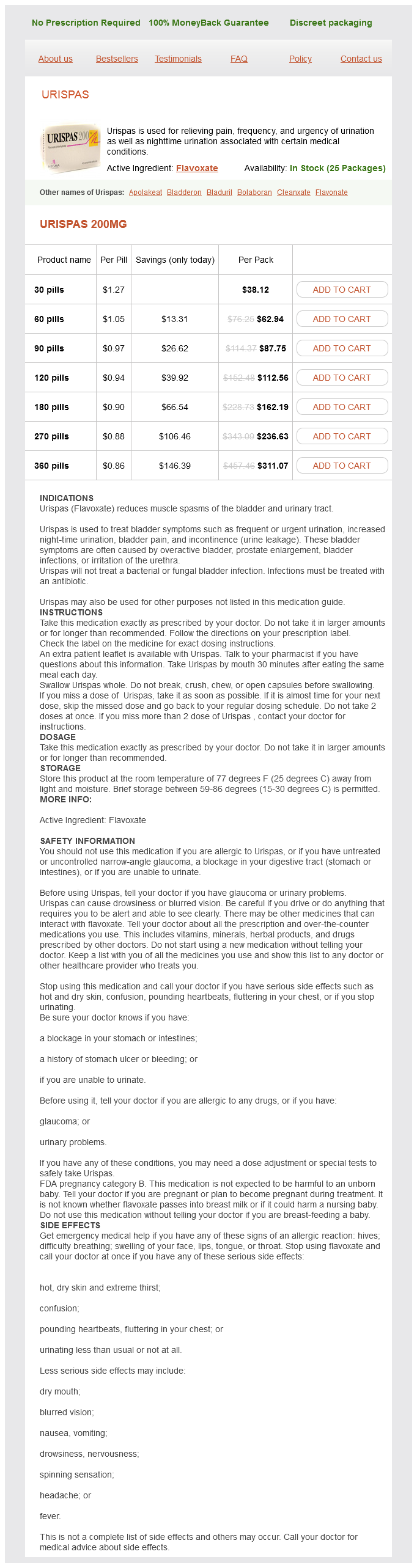
Urispas Dosage and Price
Urispas 200mg
- 30 pills - $38.12
- 60 pills - $62.94
- 90 pills - $87.75
- 120 pills - $112.56
- 180 pills - $162.19
- 270 pills - $236.63
- 360 pills - $311.07
The clear spaces around the neurons and their processes are artifacts caused by the chemical preparations of the nervous tissue spasms pancreas generic urispas 200 mg. Within the cytoplasm of the motor neurons (1) is the cytoskeleton consisting of numerous neurofibrils (3) that course through the cell body and extend into the dendrites (4) and axons (8). Visible also are numerous axons of a size different from other nerve cells in the spinal cord. Surrounding the motor neurons (1) are nuclei of neuroglial cells (2), a blood vessel (7) with blood cells and a meshwork of neural processes, the neuropil (9). Although there are variations in the arrangement of cells in the cerebral cortex, distinct layers are recognized in most regions. Horizontal and radial axons associated with neuronal cells in different layers give the cerebral cortex a laminated appearance. Overlying the molecular cell layer (I) is the delicate connective tissue of the brain, the pia mater (1). The peripheral portion of molecular layer (I) is composed predominantly of 350 neuroglial cells (2) and horizontal cells of Cajal. Their axons contribute to the horizontal fibers that are seen in the molecular layer (I). The pyramidal cells get progressively larger in successively deeper layers of the cortex. The apical dendrites of the pyramidal cells (4, 7) are directed toward the periphery of the cortex, whereas their axons extend from the cell bases. The internal pyramidal layer (V) contains neuroglial cells and the largest pyramidal cells (8), especially in the motor area of the cerebral cortex. The most prominent cell processes are the apical dendrites (1, 7) of the pyramidal cells (3), which are directed toward the surface of the cortex. The axons (4, 10) of the pyramidal cells (3) arise from the base of the cell body and pass into the white matter. The intercellular area is occupied by neuroglial cells (2, 8) in the cortex, small astrocytes, and blood vessels-venule (5) and capillary (6). The cerebellar folia (6) are covered by the thin connective tissue, the pia mater (7), which follows the surface of each folium (6) into the adjacent sulci (9). The detachment of the pia mater (7) from the cerebellar cortex (1, 10) is an artifact caused by tissue fixation and preparation. Three distinct cell layers are distinguished in the cerebellar cortex (1, 10): an outer molecular layer (2) with few and small neuronal cell bodies and fibers that extend parallel to the length of the folium, a central or middle Purkinje cell layer (3), and an inner granular layer (4) with small neurons that exhibit stained nuclei. The Purkinje cells (3) are pyriform, or pyramidal, in shape with ramified dendrites that extend into the molecular layer (2). The white matter (5, 8) forms the core of each cerebellar folium (6) and consists of myelinated nerve fibers, or axons. The Purkinje cells (3) form the Purkinje cell layer (7), with their prominent nuclei and nucleoli, and are arranged in a single row between the molecular cell layer (6) and the granular cell layer (4). The large "flask-shaped" bodies of the Purkinje cells (3, 7) exhibit thick dendrites 353 (2) that branch throughout the molecular cell layer (6) to the cerebellar surface. Thin axons (not shown) leave the base of the Purkinje cells, pass through the granular cell layer (4), become myelinated, and enter the white matter (5, 11). The molecular cell layer (6) contains basket cells (1) with unmyelinated axons that course horizontally. Descending collaterals of more deeply placed basket cells (1) arborize around the Purkinje cells (3, 7). Axons of the granule cells (9) in the granular cell layer (4) extend into the molecular layer (6) and also course horizontally as unmyelinated axons. Throughout the granular layer are small, irregularly dispersed, clear spaces called the glomeruli (10) that contain only synaptic complexes. The fibrous astrocytes (2, 5) exhibit a small cell body (5), a large oval nucleus (5), and a dark-stained nucleolus (5). Extending from the cell body are long, thin, and smooth radiating processes (4, 6) found between the neurons and blood vessels. A perivascular fibrous astrocyte (2) surrounds a capillary (8) with red blood cells (erythrocytes). From other fibrous astrocytes (2, 5), the long processes (4, 6) extend to and terminate on the capillary (8) as perivascular endfeet (3, 7). Also seen in the illustration are nuclei of different neuroglial (1) cells of the brain. Lining the capillary lumen is a thin endothelial layer and the nucleus of an endothelial cell (2). Attached externally to the capillary wall (5) are numerous perivascular endfeet of astrocytes (3, 4) that completely envelop the capillary wall (5) to form part of the bloodbrain barrier. In comparison to a fibrous astrocyte (3), the oligodendrocytes (1, 4, 7) are smaller and exhibit few, thin, short processes without excessive branching. Two neurons (2, 6) are also illustrated to contrast their size with those of a fibrous astrocyte (3) and the oligodendrocytes (1, 4, 7). The cytoplasm exhibits a well-developed granular endoplasmic reticulum (3, 5), a Golgi apparatus (6), and numerous free ribosomes around the organelles. Numerous myelinated axons (1, 4, 8), cut in cross and longitudinal sections, are surrounded with myelin sheaths (7) that are associated with the cytoplasm of the oligodendrocyte. Located in the myelinated axons (1, 4) are oval, dark-staining mitochondria (4) and numerous neurofilaments (8).